What Are Neuroendocrine Tumors (NETs)?
When you first hear that you've got a neuroendocrine tumor, you'll have lots of questions about what it is and how it will affect you. There are quite a few types of this disease, and it can show up in many places in your body.
Your symptoms may depend on where your tumor is growing and what kind it is. Learn as much as you can about your own type of NET, so you can be a confident partner with your doctor when you make decisions on a treatment plan.
While all this is going on, don't neglect your emotional needs. Your doctor can tell you how to find a support group where you can talk to others who are going through the same things you are. And feel free to open up to your friends and family about how you're doing. They know you best and can be a huge source of support.
The first thing you want to find out about your condition is where your tumor is located. NETs grow in cells that make hormones -- chemicals that help control different actions in your body, like hair growth, your sex drive, and even your mood. A neuroendocrine tumor can grow in spots like your pancreas, a gland in your belly. It can also happen in your stomach, intestines, or lungs.
Some NETs are cancer, which means they can spread to other parts of your body. Many of these tumors also make hormones of their own, which can give you certain symptoms. Other kinds of neuroendocrine tumors are benign, which means they don't move from their original spot.
Most neuroendocrine tumors grow slowly -- over years, not months -- compared with other types of tumors. Often, doctors can remove or shrink them with different treatments. Other therapies can make your symptoms better.
There are many types of NETs. They're usually named after the type of cell where they grow, or the hormone they make.
Carcinoid tumors can form in many areas of your body, but they're most common in the cells of the digestive system -- the stomach, small intestines, appendix, and rectum. They can also form in the lungs or a small organ behind the breast bone called the thymus. More rarely, they grow in the pancreas, kidneys, ovaries, or testicles.
These tumors can release different types of hormones, which can affect how you feel. Doctors call these groups of symptoms carcinoid syndrome.
Pancreatic NETs grow in your pancreas. There are a few kinds of them:
Insulinomas are the most common type. Their cells make insulin, the hormone that controls blood sugar levels. Most of the time, they're not cancerous.
Glucagonomas make glucagon, a hormone that raises your blood sugar level. About half of them are cancerous, and they often spread to other parts of your body.
Gastrinomas make the hormone gastrin, which helps you digest food. These tumors can happen if you have a rare disorder called Zollinger-Ellison syndrome. About half of these gastrinomas are cancerous, and they often spread easily in the body.
Somatostatinomas make too much of a chemical called somatostatin that affects how your body makes other hormones.
VIPomas make a hormone that triggers the release of other hormones, called vasoactive intestinal peptides (VIP). Most VIPomas are cancerous.
Some other types of NETs include:
Medullary carcinoma. It shows up in your thyroid gland, an organ that's on the base of your neck. This tumor grows in cells that make a hormone that affects the levels of calcium in your body.
Pheochromocytoma. This grows in cells of your adrenal glands, which sit above your kidneys. It makes the hormones adrenaline and noradrenaline, which can increase your heart rate and blood pressure. Usually these tumors are not cancerous.
Causes
Most of the time, doctors don't know what causes NETs. But you're more likely to get them if you have certain diseases that run in your family, such as:
Multiple endocrine neoplasia type 1. This causes tumors to grow in the pancreas and other organs.
Neurofibromatosis type 1. This can cause tumors in your adrenal glands.
Von Hippel-Lindau syndrome. It makes tumors and fluid-filled sacs form in many parts of your body.
Symptoms
How a neuroendocrine tumor makes you feel depends on the type you have and where it is in your body.
With a pancreatic NET, you might have:
Blurred or double vision
Confusion
Diarrhea
Dizziness
A fast heartbeat
Headache
Hunger that's stronger than usual
Rash
Shakiness
Stomach pain
Sweating
Weakness
Weight loss without trying
Carcinoid tumors can cause:
Diarrhea
Red, warm, itchy skin, often on your face and neck
Coughing
Pain in your chest
Stomach pain
Feeling tired or sick
Trouble breathing
Weight gain or loss without trying
Other types of NETs can cause:
Appetite loss
Bleeding
Coughing
Diarrhea
Fever
Headache
A hoarse voice
A fast heart rate
Nausea or vomiting
Night sweats
Pain
Rash
Sweating
Weight gain or loss without trying
Yellowish skin or eyes
Getting a Diagnosis
When you see your doctor, he'll give you a physical exam, and he'll want to hear about how you're feeling. He might ask you questions like:
How long have you been feeling this way?
Do you have any pain? Where?
How is your appetite?
Have you gained or lost any weight?
Do you feel weak or more tired than usual?
Do you have any skin rashes?
Do you have any medical conditions?
Are there any illnesses that run in your family?
Your doctor can use a few different tests to check for a tumor in your body. You might get:
Blood and urine tests. They check the levels of hormones in your body to see if they're too high or too low.
CT scan. It's a powerful X-ray that makes detailed pictures inside your body.
MRI. It uses powerful magnets and radio waves to make pictures of your organs.
Octreotide scan. In a hospital, you'll get a shot of a small amount of a radioactive liquid through an IV. Then, you'll lie down in a scanner that can make images of your insides. The liquid has a drug called octreotide that will stick to cells on the surface of most NETs. The radiation in the fluid helps doctors see those cells on the picture from the scanner. You'll get two scans over 2 days, but you won't have to spend the night in the hospital. Each scan can take up to 3 hours, but it won't hurt.
X-ray. It uses radiation in low doses to show the inside of your body.
Biopsy. Your doctor will take a small piece of tissue from your body and look at it under a microscope to check for tumor cells. He may use a CT scan to help him find the right area. Or he may use a thin, flexible tube with a small camera, called an endoscope, to look at the lining of your digestive tract. You might be asleep or awake during the procedure, but you'll get medicine to make you more comfortable.
Molecular testing. Your doctor checks the sample of the tumor from the biopsy for certain genes, proteins, and other substances. The results help him decide what kind of treatment you need.
Questions for Your Doctor
What type of NET do I have, and where is it? Is it cancerous?
What does this mean for me?
Have you treated people with this kind of NET before?
Is surgery an option for me?
What other treatments do you recommend?
How will they make me feel?
How will we know if it's working?
What changes should I expect in my daily life?
Will my children get a NET, too?
Treatment
Doctors can treat NETs with surgery, radiation, chemotherapy, and drugs. The treatment you get will depend on:
What kind of tumor you have and how many there are
Whether it's cancerous
If it has spread to other parts of your body
Brain Tumors
Types of Brain Tumors
A brain tumor, known as an intracranial tumor, is an abnormal mass of tissue in which cells grow and multiply uncontrollably, seemingly unchecked by the mechanisms that control normal cells. More than 150 different brain tumors have been documented, but the two main groups of brain tumors are termed primary and metastatic.
Primary brain tumors include tumors that originate from the tissues of the brain or the brain's immediate surroundings. Primary tumors are categorized as glial (composed of glial cells) or non-glial (developed on or in the structures of the brain, including nerves, blood vessels and glands) and benign or malignant.
Metastatic brain tumors include tumors that arise elsewhere in the body (such as the breast or lungs) and migrate to the brain, usually through the bloodstream. Metastatic tumors are considered cancer and are malignant.
Metastatic tumors to the brain affect nearly one in four patients with cancer, or an estimated 150,000 people a year. Up to 40 percent of people with lung cancer will develop metastatic brain tumors. In the past, the outcome for patients diagnosed with these tumors was very poor, with typical survival rates of just several weeks. More sophisticated diagnostic tools, in addition to innovative surgical and radiation approaches, have helped survival rates expand up to years; and also allowed for an improved quality of life for patients following diagnosis.
Types of Benign Brain Tumors
Chordomas are benign, slow-growing tumors that are most prevalent in people ages 50 to 60. Their most common locations are the base of the skull and the lower portion of the spine. Although these tumors are benign, they may invade the adjacent bone and put pressure on nearby neural tissue. These are rare tumors, contributing to only 0.2 percent of all primary brain tumors.
Craniopharyngiomas typically are benign, but are difficult tumors to remove because of their location near critical structures deep in the brain. They usually arise from a portion of the pituitary gland (the structure that regulates many hormones in the body), so nearly all patients will require some hormone replacement therapy.
Gangliocytomas, gangliomas and anaplastic gangliogliomas are rare tumors that include neoplastic nerve cells that are relatively well-differentiated, occurring primarily in young adults.
Glomus jugulare tumors most frequently are benign and typically are located just under the skull base, at the top of the jugular vein. They are the most common form of glomus tumor. However, glomus tumors, in general, contribute to only 0.6 percent of neoplasms of the head and neck.
Meningiomas are the most common benign intracranial tumors, comprising 10 to 15 percent of all brain neoplasms, although a very small percentage are malignant. These tumors originate from the meninges, the membrane-like structures that surround the brain and spinal cord.
Pineocytomas are generally benign lesions that arise from the pineal cells, occurring predominantly in adults. They are most often well-defined, noninvasive, homogeneous and slow-growing.
Pituitary adenomas are the most common intracranial tumors after gliomas, meningiomas and schwannomas. The large majority of pituitary adenomas are benign and fairly slow-growing. Even malignant pituitary tumors rarely spread to other parts of the body. Adenomas are by far the most common disease affecting the pituitary. They commonly affect people in their 30s or 40s, although they are diagnosed in children, as well. Most of these tumors can be treated successfully.
Schwannomas are common benign brain tumors in adults. They arise along nerves, comprised of cells that normally provide the "electrical insulation" for the nerve cells. Schwannomas often displace the remainder of the normal nerve instead of invading it. Acoustic neuromas are the most common schwannoma, arising from the eighth cranial nerve, or vestibularcochlear nerve, which travels from the brain to the ear. Although these tumors are benign, they can cause serious complications and even death if they grow and exert pressure on nerves and eventually on the brain. Other locations include the spine and, more rarely, along nerves that go to the limbs.
Types of Malignant Brain Tumors
Gliomas are the most prevalent type of adult brain tumor, accounting for 78 percent of malignant brain tumors. They arise from the supporting cells of the brain, called the glia. These cells are subdivided into astrocytes, ependymal cells and oligodendroglial cells (or oligos). Glial tumors include the following:
Astrocytomas are the most common glioma, accounting for about half of all primary brain and spinal cord tumors. Astrocytomas develop from star-shaped glial cells called astrocytes, part of the supportive tissue of the brain. They may occur in many parts of the brain, but most commonly in the cerebrum. People of all ages can develop astrocytomas, but they are more prevalent in adults — particularly middle-aged men. Astrocytomas in the base of the brain are more prevalent in children or younger people and account for the majority of children's brain tumors. In children, most of these tumors are considered low-grade, while in adults, most are high-grade.
Ependymomas are derived from a neoplastic transformation of the ependymal cells lining the ventricular system and account for two to three percent of all brain tumors. Most are well-defined, but some are not.
Glioblastoma multiforme (GBM) is the most invasive type of glial tumor. These tumors tend to grow rapidly, spread to other tissue and have a poor prognosis. They may be composed of several different kinds of cells, such as astrocytes and oligodendrocytes. GBM is more common in people ages 50 to 70 and are more prevalent in men than women.
Medulloblastomas usually arise in the cerebellum, most frequently in children. They are high-grade tumors, but they are usually responsive to radiation and chemotherapy.
Oligodendrogliomas are derived from the cells that make myelin, which is the insulation for the wiring of the brain.
Other Types of Brain Tumors
Hemangioblastomas are slow-growing tumors, commonly located in the cerebellum. They originate from blood vessels, can be large in size and often are accompanied by a cyst. These tumors are most common in people ages 40 to 60 and are more prevalent in men than women.
Rhabdoid tumors are rare, highly aggressive tumors that tend to spread throughout the central nervous system. They often appear in multiple sites in the body, especially in the kidneys. They are more prevalent in young children, but also can occur in adults.
Brain Cancer
Brain tumours include types of brain cancer, however not all brain tumours are cancerous.
Brain tumours are graded 1-4 by their behaviour such as speed of growth and how likely they are to spread. These grades are then split into low grade (1-2) and high grade (3-4), with low grade tumours defined as non-cancerous and high grade tumours as cancerous.
However, it is important to remember that just because a tumour is low grade, it does not mean there are no associated health risks or problems. Having regular check-ups is important whether you have a high or low grade tumour.
What is brain cancer?
Following the diagnosis of brain cancer, the first question you may ask is 'what is brain cancer'?
We have billions of cells in our body, which grow and multiply to help support our body's natural processes and functions, such as repairing damage.
However, if the cells in the brain 'go wrong' and begin to grow in an abnormal way, rather than repair damage, they can inadvertently cause it.
If these abnormal brain cells begin to grow and multiply, contained within the brain, this is how a primary brain tumour can occur. If the cells then grow rapidly and spread within the brain, this is how cancerous tumours are formed and will result in a brain cancer diagnosis.
If the cells go wrong elsewhere in the body first, say, the lungs, and those cells spread to the brain, this is known as secondary brain cancer or, metastases.
How brain cancer is graded
There are over 150 different brain tumour types, each named after the type of cell they grow from, their location in the brain how likely they are to spread.
Brain tumours that grow rapidly are known as high grade (grade 3 brain cancer and grade 4 brain cancer).
Occasionally, people will refer to these as stage 3 brain cancer or stage 4 brain cancer. However the word 'stage' is often used in other forms of cancer, but is incorrect when discussing brain cancer.
Brain tumours that grow more slowly, and are usually non-cancerous, are known as low grade (grade 1 brain tumour and grade 2 brain tumour).
The prognosis for brain cancer varies from person to person and your medical team is best placed to advice you of this based on your individual circumstances and brain cancer diagnosis.
What is the cause of brain cancer
It is important to remember that there is nothing you could have done, or not done, to prevent brain cancer.
Largely, there is no known cause of brain cancer, but we do know there are risk factors, such as your genetic makeup or exposure to radiation.
Inheriting a gene which makes you more likely to develop brain cancer: it is estimated that an inherited gene accounts for one in 20 cases of brain tumours. Certain genetic conditions may also increase your risk of developing a low or high grade brain tumour.
Exposure to radiation: the risk of some brain tumour types (meningioma or glioma) is higher if you had radiotherapy to your head as a child, this is particularly the case if it occurred before the age of five.
Can brain cancer be cured?
Many people diagnosed with brain cancer will want to know if brain cancer be cured, however this can vary from person to person and type to type.
It is more likely that brain cancer will spread to other parts of the brain than a lower grade brain tumour and, despite successful treatment, brain cancer can often return.
However, this depends on a lot of factors such as the location of the tumour, its reaction to treatment, or the success of surgery and, to a certain extent, its molecular/genetic make-up. Your medical team will be best placed to advice you on your individual circumstances and prognosis.
Types of brain cancer
The most common type of primary brain cancer in adults is glioblastoma.
There are both primary and secondary types of glioblastoma. Primary glioblastoma originates in the brain and first appears as a grade 4 glioblastoma.
Secondary brain cancer
Often, secondary cancer refers to the spread of cancer from one part of the body to another, however a secondary glioblastoma still originates in the brain but has developed from a lower grade brain tumour type, known as an astrocytoma.
Brain Cancer Prognosis
The prognosis for brain cancer types varies from type to type and person to person and depends on a lot of factors, such as the location of the tumour, its reaction to treatments, or the success of surgery. Your medical team will be best placed to advice you based on your individual circumstances and your brain cancer diagnosis.
If you brain cancer type is classed as inoperable this means that your medical team may not be able to perform surgery for reasons such as the location of the tumour. For example if it is too close to vital structures of the brain, or because the cancer is not solid lump or mass and it is therefore difficult to identify the edges of the tumour. Operating in such circumstances could result in damage to healthy brain tissue and vital areas of the brain that control movement, sight or breathing.

CT scans, commonly used in medical imaging, may increase the risk of brain tumours, a study has found.
The use of computed tomography (CT) scans has increased dramatically over the last two decades. CT scans greatly improve diagnostic capabilities, which in turn improve clinical outcomes.
However, they deliver higher radiation doses, and can especially affect children who are more susceptible to radiation-related malignancies than adults, researchers said.
The most common malignancies caused by radioactivity among children and young adults are leukaemia and brain tumours.
Researchers from Netherlands Cancer Institute evaluated leukaemia and brain tumour risks following exposure to radiation from CT scans in childhood.
For a group of 168,394 Dutch children who received one or more CT scans between 1979 and 2012, researchers obtained cancer incidence and vital status by record linkage.
They surveyed all Dutch hospital-based radiology departments to ascertain eligibility and participation. In the Netherlands, paediatric CT scans are only performed in hospitals.
Overall cancer incidence was 1.5 times higher than expected. For all brain tumours combined, and for malignant and nonmalignant brain tumours separately, dose-response relationships were observed with radiation dose to the brain.
Relative risks increased to between two and four for the highest dose category. The researchers observed no association for leukaemia. Radiation doses to the bone marrow, where leukaemia originates, were low.
They caution that this pattern of excess cancer risk may be partly due to confounding by indication because the incidence of brain tumours was higher in the cohort than in the general population.
CT scans are sometimes used to identify conditions associated with an increased tumour risk; the reason these children had CT scans may be associated with their risk of developing cancer.
"Epidemiological studies of cancer risks from low doses of medical radiation are challenging, said the study's principal investigator, Michael Hauptmann, from Netherlands Cancer Institute.
"Our careful evaluation of the data and evidence from other studies indicate that CT-related radiation exposure increases brain tumour risk," said Hauptmann.

न्यूरोएन्डोक्राइन ट्यूमर म्हणजे ब्रेन टय़ूमर
‘ब्रेन टय़ूमर झाला की सगळे संपले!’ अशी अनेकांची भूमिका असते. ‘टय़ूमर म्हणजे कॅन्सरच’ अशा भ्रमातही अनेकजण असतात. या चुकीच्या समजामुळे ब्रेन टय़ूमर हा ‘भीतीचा गोळा’ ठरतो! ब्रेन टय़ूमर म्हणजे नेमके काय, तो कसा होतो, त्यावरील अत्याधुनिक उपचारपद्धती कोणत्या, याबद्दल माहिती घेऊया या लेखामधून..
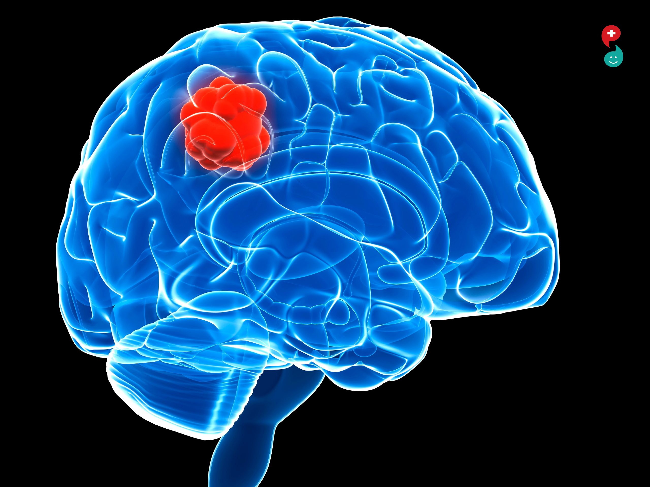
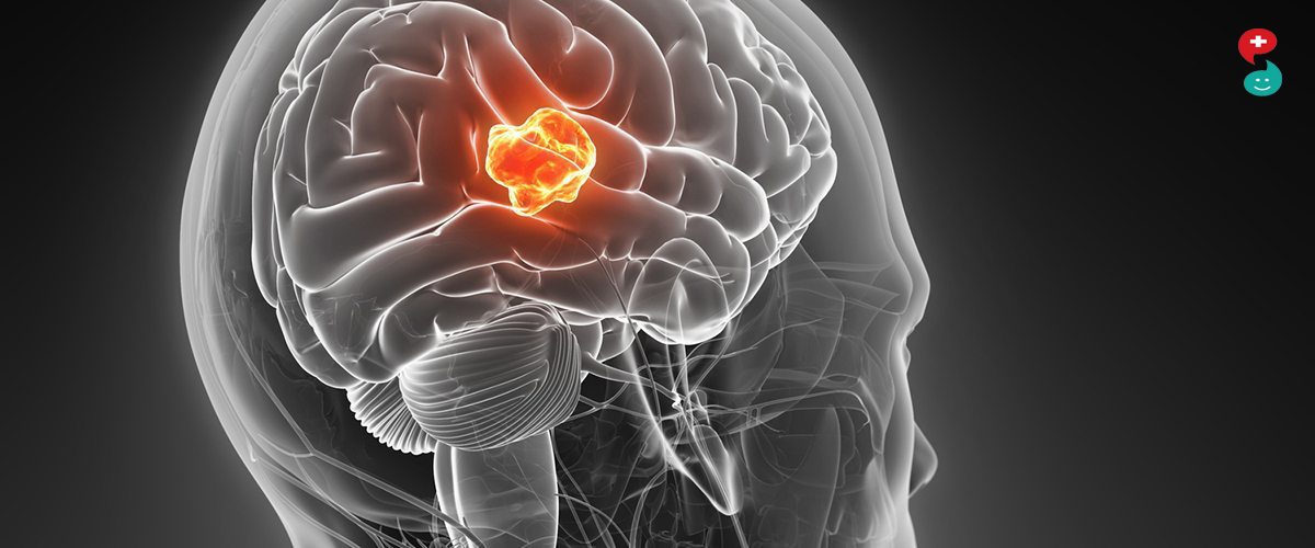
बंद कवटीत जेव्हा ब्रेन टय़ूमरची गाठ जागा व्यापू लागते तेव्हा तिचा दाब मेंदूवर पडायला लागतो. त्यामुळे ब्रेन टय़ूमरच्या ७५ ते ८० टक्के रुग्णांमध्ये टय़ूमर झाल्याची लक्षणे दिसून येतात. डोकेदुखी, उलटी होणे अशा लक्षणांबरोबरच मेंदूच्या ज्या भागावर दाब पडतो त्या भागाशी संबंधित असलेल्या शारीरिक कार्यात बिघाड होणे अशी लक्षणे दिसू शकतात. या प्रकारात दृष्टी अधू होणे, दृष्टीस पडलेल्या गोष्टी लक्षात न राहणे, रंग ओळखता न येणे, बोलताना अडखळणे, हाता-पायातली ताकद कमी होणे अशा तक्रारीही आढळतात, पण वीस टक्के रुग्णांमध्ये कोणतीही लक्षणे दिसून येत नाहीत. काही रुग्णांत ब्रेन टय़ूमर खूप सावकाश वाढतो. त्यामुळे मेंदूतली जागा व्यापली जाण्याची प्रक्रियाही सावकाश होते. अशा रुग्णांमध्ये मेंदूला या गाठीची सवय होत जाते आणि त्यामुळे कोणतीही लक्षणेही दिसत नाहीत.
‘टय़ूमर झाला म्हणजे तो कॅन्सरच असणार’, असा सार्वत्रिक समज आपल्याकडे आढळून येतो, हे मात्र खरे नाही. टय़ूमरची गाठ कॅन्सरची नसलेलीही असू शकते हे लक्षात घेणे आवश्यक आहे. मेंदूतील टय़ूमरची गाठ कॅन्सरची असण्याची शक्यता साधारणपणे ५० ते ६० टक्के असते. म्हणजेच उरलेल्या ५० टक्के रुग्णांचा टय़ूमर हा कॅन्सर नसतो. मेंदूतील कॅन्सरच्या नसलेल्या गाठी शस्त्रक्रियेद्वारे पूर्णपणे बाहेर काढता येतात. शस्त्रक्रियेनंतर रुग्ण अल्पावधीत बरा होऊन अगदी पूर्वीसारखे आयुष्यही जगू लागतो. हे बऱ्याच रुग्णांना माहिती नसल्यामुळे ‘टय़ूमर झाला म्हणजे सगळे संपले’ या भावनेने रुग्ण आधीच मनाने खचतात. ‘टय़ूमरची शस्त्रक्रिया म्हणजे मरण’ या चुकीच्या कल्पनेमुळे काही रुग्ण शस्त्रक्रियेलाच नकार देऊन ‘असेल-नसेल ते आयुष्य तरी बरे जगावे!’ अशी भूमिका घेतात. त्यामुळे रुग्णांनी आपल्याला काय झाले आहे याविषयी वाचनाद्वारे माहिती मिळवत राहणे आवश्यक आहे. त्याद्वारे त्यांची दिशाभूल होणे टळेल.
गेल्या तीस वर्षांत ब्रेन टय़ूमरच्या शस्त्रक्रियांमध्ये प्रचंड प्रमाणात बदल झाले आहेत. पूर्वी जे टय़ूमर असाध्य मानले जात ते आज साध्य आहेत. ज्या शस्त्रक्रिया करायला पूर्वी दहा-दहा तास लागत त्या शस्त्रक्रिया आता दोन तासांतही होऊ शकतात. एमआरआय प्रतिमांसारख्या तंत्रांद्वारे टय़ूमरची गाठ खूप लहान असतानाच लक्षात येते. शस्त्रक्रिया झाल्यानंतर दहा मिनिटांत रुग्ण शुद्धीवर येतो, बोलायलाही लागतो! लहान शस्त्रक्रियेनंतर तर रुग्णाला दुसऱ्या दिवशी रुग्णालयातून घरी सोडणेही शक्य होते. हा आमूलाग्र बदल गेल्या दहा वर्षांत झाला आहे.
परदेशात ज्या अत्याधुनिक शस्त्रक्रिया केल्या जातात त्याही आज मुंबई-पुण्यात यशस्वीपणे केल्या जाऊ शकतात, पण राज्यातील जवळजवळ सर्वच जिल्ह्य़ांत ब्रेन टय़ूमरच्या शस्त्रक्रिया होण्याइतपत सुविधा नक्कीच उपलब्ध आहेत. मेंदूत रक्ताची गुठळी झालेल्या रुग्णालाही वाचवणे पूर्वी अवघड होत असे. आता तालुका पातळीवरही या शस्त्रक्रिया होऊ लागल्या आहेत. अद्ययावत दुर्बिणींचा शस्त्रक्रियेसाठी वापर, ‘क्यूसा’ म्हणजे ‘कॅव्हिट्रॉन अल्ट्रासाऊंड अॅस्पिरेटर’ चा वापर यामुळे शस्त्रक्रिया अधिक सुकर बनल्या आहेत. टय़ूमरचे लहान तुकडे करून काढण्याऐवजी क्यूसा या यंत्रणेद्वारे टय़ूमरला ‘व्हेपराईज’ करून म्हणजे वाफ स्वरूपात आणून शोषून घेता येते. आता ‘स्टिरिओटॅक्टिक सर्जरी’ त मेंदूतील कोणत्याही भागापर्यंत अचूकतेने पोहोचून टय़ूमरची ‘बायोप्सी’ करता येते. यात सर्वच रुग्णांना पूर्ण भूल देण्याचीही गरज नसते. ‘लोकल अॅनास्थिशिया’ देऊनही बायोप्सी करता येते. रुग्ण एकीकडे बोलत असताना त्याच्या मेंदूत सरळ ‘प्रोब’ घालून टय़ूमरची बायोप्सी काढणे म्हणजे पुढील निदानासाठी टय़ूमरचा तुकडा किंवा पस काढणे शक्य होते. या तुकडय़ाचा दुर्बिणीखाली अभ्यास केला जातो. त्यात कोणत्या प्रकारच्या पेशी आहेत यावर ती गाठ साधी आहे की कॅन्सरची आहे, याचे निदान होते. त्यावर पुढील उपचारांची दिशा ठरते.
मानवी जनुकांमध्ये होणारे बदल टय़ूमरसाठी मूलत: कारणीभूत ठरतात. प्रत्येक पेशीत आपल्यासारखीच पेशी निर्माण करण्याचा गुणधर्म असतो. ही नवी पेशी जुन्या पेशीसारखीच असते. त्यामुळेच शरीरातील काही पेशी खराब झाल्या तरी त्यांची कमतरता नव्या पेशी भरून काढतात. जनुकावर विपरीत परिणाम झाला तर नव्याने तयार होणारी पेशी वेगळ्या प्रकारची असू शकते. अशा वेगळ्या पेशींपासूनच पुढे गाठ निर्माण होण्याची शक्यता असते. भोवतालच्या नैसर्गिक बदलांचे दूरगामी परिणामही जनुकीय बदलांमध्ये दिसू शकतात. मोबाईल फोनचा अतिवापर ब्रेन टय़ूमरला कारणीभूत ठरतो असे म्हटले जाते. हे पुराव्यानिशी जरी सिद्ध करता येत नसले तरी प्रबळ इलेक्ट्रोमॅग्नेटिक लहरी मानवातील जनुकीय बदलांसाठी कारणीभूत ठरू शकतात.
‘अमुक एक गोष्ट केली तर ब्रेन टय़ूमर होईल’, आणि ‘अमुक एक केले तर तो टाळता येईल’, अशी सोपी विभागणी करणे अशक्य आहे. पण ब्रेन टय़ूमर झाल्यानंतरही जीवन असाध्य नक्कीच नाही! त्यातून वाट काढायचे मार्ग आहेत. ते अगदी अत्याधुनिक आणि यशस्वितेच्या कसोटीवर उतरलेले आहेत. उपचारांनंतर रुग्ण त्याचे उरलेले आयुष्य अगदी पुर्वीसारखेच जगू शकतो. त्यामुळे ब्रेन टय़ूमर रुग्णाच्या आयुष्यात एखाद्या परीक्षेसारखा असेल कदाचित् पण तो भीतीचा गोळा ठरायला नको!