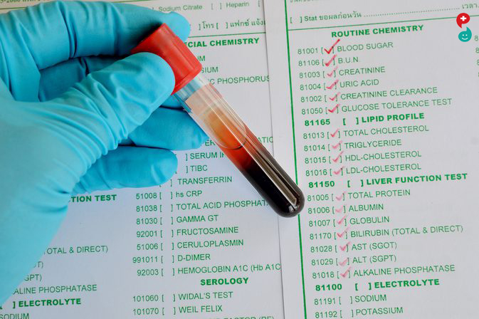
Chloride is an electrolyte. It is a negatively charged ion that works with other electrolytes, such as potassium, sodium, and bicarbonate, to help regulate the amount of fluid in the body and maintain the acid-base balance. This test measures the level of chloride in the blood and/or urine.
Chloride is present in all body fluids but is found in the highest concentration in the blood and in the fluid outside of the body's cells. Most of the time, chloride concentrations mirror those of sodium, increasing and decreasing for the same reasons and in direct relationship to sodium. When there is an acid-base imbalance, however, blood chloride levels can change independently of sodium levels as chloride acts as a buffer. It helps to maintain electrical neutrality at the cellular level by moving into or out of the cells as needed.
We get chloride in our diet through food and table salt, which is made up of sodium and chloride ions. Most of the chloride is absorbed by the digestive tract, and the excess is eliminated in urine. The normal blood level remains steady, with a slight drop after meals (because the stomach produces acid after eating, using chloride from blood).
A chloride blood test is used to detect abnormal concentrations of chloride. It is often ordered, along with other electrolytes, as part of a regular physical to screen for a variety of conditions.
Chloride is an electrolyte. It is a negatively charged ion that works with other electrolytes, such as potassium, sodium, and bicarbonate, to help regulate the amount of fluid in the body and maintain the acid-base (pH) balance.
Chloride and electrolyte tests may also be ordered to help diagnose the cause of signs and symptoms such as prolonged vomiting, diarrhea, weakness, and difficulty breathing (respiratory distress). If an electrolyte imbalance is detected, a healthcare practitioner will look for and address the disease, condition, or medication causing the imbalance and may order electrolyte testing at regular intervals to monitor the effectiveness of treatment. If an acid-base imbalance is suspected, the healthcare practitioner may also order tests for blood gases to further evaluate the severity and cause of the imbalance.
In persons with too much base, urine chloride measurements can tell the healthcare practitioner whether the cause is loss of salt (in cases of dehydration, vomiting, or use of diuretics, where urine chloride would be very low) or an excess of certain hormones such as cortisol or aldosterone that can affect electrolyte elimination.
What is being tested?
Chloride is an electrolyte. It is a negatively charged ion that works with other electrolytes, such as potassium, sodium, and bicarbonate, to help regulate the amount of fluid in the body and maintain the acid-base balance. This test measures the level of chloride in the blood and/or urine.
What are catecholamines?
The catecholamine blood test measures the amount of catecholamines in your body.
Catecholamines is an umbrella term for the hormones dopamine, norepinephrine, and epinephrine, which naturally occur in your body.
Doctors usually order the test to check for adrenal tumors in adults. These are tumors that affect the adrenal gland, which sits on top of the kidney. The test also checks for neuroblastomas, a cancer that starts in the sympathetic nervous system, in children.
Your body produces more catecholamines during times of stress. These hormones prepare your body for stress by making your heart beat faster and raising your blood pressure.
What is the purpose of the catecholamine blood test?
The catecholamine blood test determines whether the level of catecholamines in your blood is too high.
Most likely, your doctor has ordered a catecholamine blood test because they're concerned that you might have a pheochromocytoma. This is a tumor that grows on your adrenal gland, where catecholamines are released. Most pheochromocytomas are benign, but it's important to remove them so they don't interfere with regular adrenal function.
Your child and the catecholamine blood test
Your child's doctor may order a catecholamine blood test if they're concerned that your child may have neuroblastoma, which is a common childhood cancer. According to the American Cancer Society, 6 percent of cancers in children are neuroblastomas. The sooner a child with neuroblastoma is diagnosed and begins treatment, the better their outlook.
What symptoms might make my doctor order a catecholamine blood test?
Symptoms of pheochromocytoma
The symptoms of a pheochromocytoma, or adrenal tumor, are:
high blood pressure
rapid heartbeat
an unusually hard heartbeat
heavy sweating
severe headaches off and on for an extended period
pale skin
unexplained weight loss
feeling unusually frightened for no reason
feeling strong, unexplained anxiety
Symptoms of neuroblastoma
The symptoms of neuroblastoma are:
painless lumps of tissue under the skin
abdominal pain
chest pain
back pain
bone pain
swelling of the legs
wheezing
high blood pressure
rapid heartbeat
diarrhea
bulging eyeballs
dark areas around the eyes
any changes to the shape or size of eyes, including changes to pupil size
fever
unexplained weight loss
powered by Rubicon Project
How to prepare and what to expect
Your doctor may tell you not to eat or drink anything for 6 to 12 hours before the test. Follow your doctor's orders carefully to ensure accurate test results.
A healthcare provider will take a small sample of blood from your veins. They'll probably ask you to remain quietly seated or to lie down for as long as half an hour before your test.
A healthcare provider will tie a tourniquet around your upper arm and look for a vein large enough to insert a small needle into. When they've located the vein, they'll clean the area around it to make sure they don't introduce germs into your bloodstream. Next, they'll insert a needle connected to a small vial. They'll collect your blood in the vial. This could sting a little. They'll send the collected blood to a diagnostic lab for an accurate reading.
Sometimes the healthcare provider taking your blood sample will access one of the veins on the back of your hand instead of inside your elbow.
What might interfere with test results?
A number of common medications, foods, and beverages can interfere with catecholamine blood test results. Coffee, tea, and chocolate are examples of things you might have recently consumed that make your catecholamine levels rise. Over-the-counter (OTC) medications, such as allergy medicine, could also interfere with the reading.
Your doctor should give you a list of things to avoid before your test. Make sure to tell your doctor all of the prescription and OTC medicines you're taking.
Since even small amounts of stress affect catecholamine levels in the blood, some people's levels may rise just because they're nervous about having a blood test.
If you're a breastfeeding mother, you may also want to check with your doctor about your intake before your child's catecholamine blood test.
What are the possible outcomes?
Because catecholamines are related to even small amounts of stress, the level of catecholamines in your body changes based on whether you're standing, sitting, or lying down.
The test measures catecholamines by picogram per milliliter (pg/mL); a picogram is one-trillionth of a gram. The Mayo Clinic lists the following as normal adult levels of catecholamines:
norepinephrine
lying down: 70750 pg/mL
standing: 2001,700 pg/mL
epinephrine
lying down: undetectable up to 110 pg/mL
standing: undetectable up to 140 pg/mL
dopamine
less than 30 pg/mL with no change in posture
Childrens levels of catecholamines vary dramatically and change by the month in some cases because of their rapid growth. Your childs doctor will know what the healthy level is for your child.
High levels of catecholamines in adults or children can indicate the presence of a neuroblastoma or a pheochromocytoma. Further testing will be necessary.
Overview
Sentinel node biopsy is a surgical procedure used to determine whether cancer has spread beyond a primary tumor into your lymphatic system. It's used most commonly in evaluating breast cancer and melanoma.
The sentinel nodes are the first few lymph nodes into which a tumor drains. Sentinel node biopsy involves injecting a tracer material that helps the surgeon locate the sentinel nodes during surgery. The sentinel nodes are removed and analyzed in a laboratory.
If the sentinel nodes are free of cancer, then cancer is unlikely to have spread, and removing additional lymph nodes is unnecessary.
If a sentinel lymph node biopsy reveals cancer, your doctor might recommend removing more lymph nodes.
Why it's done
Sentinel node biopsy is recommended for people with certain types of cancer to determine whether the cancer cells have spread into the lymphatic system.
Sentinel node biopsy is routinely used for people with:
Breast cancer
Melanoma
Sentinel node biopsy is being studied for use with other types of cancer, such as:
Colon cancer
Esophageal cancer
Head and neck cancer
Non-small cell lung cancer
Stomach cancer
Thyroid cancer
Risks
Sentinel node biopsy is generally a safe procedure. But as with any surgery, it carries a risk of complications, including:
Bleeding
Pain or bruising at the biopsy site
Infection
Allergic reaction to the dye used for the procedure
Lymphedema a condition in which your lymph vessels drain all the lymph fluid from an area of your body, causing fluid buildup and swelling
Lymphedema
Although lymphedema is an unlikely complication of sentinel node biopsy, one of the main reasons sentinel node biopsy was developed was to decrease the chance of developing lymphedema, which is more likely to occur if many lymph nodes are removed from one area.
Because only a few lymph nodes are removed, the risk of lymphedema after sentinel node biopsy is small. Dozens of other lymph nodes remain in the area of your body where the sentinel node biopsy is done. In most cases, those remaining lymph nodes can effectively process the lymph fluid.
How you prepare
Your doctor might ask you to avoid eating and drinking for a certain period of time before the procedure to avoid anesthesia complications. Ask your doctor about your situation.
What you can expect
Before the procedure
The first step in a sentinel node biopsy is to locate the sentinel nodes. Options include:
Radioactive solution. In this option, a weak radioactive solution is injected near the tumor. This solution is taken up by your lymphatic system and travels to the sentinel nodes.
This injection is usually done several hours or the day before the surgical procedure to remove the sentinel nodes.
Blue dye. Your doctor might inject a harmless blue dye into the area near the tumor. Your lymphatic system delivers the dye to the sentinel nodes, staining them bright blue.
You might notice a change in your skin color at the injection site. This color usually disappears in time, but it can be permanent. You might also notice that your urine is blue for a brief time.
The blue dye is typically injected just before the surgical procedure to remove the sentinel nodes.
Whether you receive the radioactive solution or the blue dye or both to locate the sentinel nodes is usually determined by your surgeon's preference. Some surgeons use both techniques in the same procedure.
During the procedure
You're likely to be under general anesthesia during the procedure.
The surgeon begins by making a small incision in the area over the lymph nodes.
If you've received radioactive solution before the procedure, the surgeon uses a small instrument called a gamma detector to determine where the radioactivity has accumulated and identify the sentinel nodes.
If the blue dye is used, it stains the sentinel nodes bright blue, allowing the surgeon to see them.
The surgeon then removes the sentinel nodes. In most cases, there are one to five sentinel nodes, and all are removed. The sentinel nodes are sent to a pathologist to examine under a microscope for signs of cancer.
In some cases, sentinel node biopsy is done at the same time as surgery to remove the cancer. Or, sentinel node biopsy can be done before or after surgery to remove the cancer.
After the procedure
You're moved to a recovery room where the health care team monitors you for complications from the procedure and anesthesia. If you don't have additional surgery, you'll be able to go home the same day.
How soon you can return to your regular activities will depend on your situation. Talk to your doctor.
If you have sentinel node biopsy as part of a procedure to remove the cancer, your hospital stay will be determined by the extent of your operation.
Results
If the sentinel nodes don't show cancer, you won't need other lymph node evaluation. If further treatment is needed, your doctor will use information from the sentinel node biopsy to develop your treatment plan.
If any of the sentinel nodes contain cancer, your doctor might recommend removing more lymph nodes to determine how many are affected.
In certain cases, a pathologist can examine the sentinel nodes during your procedure. If the sentinel lymph node shows cancer, you might need to have more lymph nodes removed right away rather than having another operation.
A complete semen analysis measures the quantity and quality of the fluid released during ejaculation. It evaluates both the liquid portion, called semen or seminal fluid, and the microscopic, moving cells called sperm. It is often used in the evaluation of male infertility. A shorter version of this test checks solely for the presence of sperm in semen a few months after a man has had a vasectomy to determine whether the surgery was successful.
Semen is a viscous, whitish liquid that contains sperm and the products from several glands. It is fairly thick at ejaculation but thins out, or liquefies, within 10 to 30 minutes. Sperm are reproductive cells in semen that have a head, midsection, and a tail and contain one copy of each chromosome (all of the male's genes). Sperm are motile, normally moving forward through the semen. Inside a woman's body, this property enables them to travel to and fuse with the female's egg, resulting in fertilization. Each semen sample is between 1.5 and 5.5 milliliters (about one teaspoon) of fluid, containing at least 20 million sperm per milliliter, and varying amounts of fructose (a sugar), buffers, coagulating substances, lubricants, and enzymes that are intended to support the sperm and the fertilization process.
A typical semen analysis measures:
Volume of semen
Viscosity consistency or thickness of the semen
Sperm counttotal number of sperm
Sperm concentration (density)number of sperm per volume of semen
Sperm motility percent able to move as well as how vigorously and straight the sperm move
Number or percent of normal and abnormal (defective) sperm in terms of size and shape (morphology)
Coagulation and liquefaction how quickly the semen turns from thick consistency to liquid
Fructose a sugar in semen that gives energy to sperm
pH measures acidity
Number of immature sperm
Number of white blood cells (cells that indicate infection)
Additional tests may be performed if the sperm count is low, if the sperm show decreased motility or abnormal morphology, or if the seminal fluid is found to be abnormal. These additional tests may help identify abnormalities such as the presence of sperm antibodies, abnormal hormone levels (testosterone, FSH, LH, prolactin), excessive number of white blood cells, and genetic tests for conditions that may affect fertility, such as Klinefelter syndrome, cystic fibrosis, or other chromosomal abnormality.
In some instances, imaging tests such as ultrasound, CAT scan, or MRI may be required. A biopsy of the testicle may also be needed. Sometimes a test called cryosurvival is done to see how well semen will survive for long-term storage if a couple would like to store sperm for future pregnancies.
How is the sample collected for testing?
Post-vasectomy sperm check: a semen sample is collected in a clean, wide-mouth container provided by the lab.
Infertility evaluation: Most laboratories require samples to be collected on-site as the semen needs to be examined within 60 minutes after ejaculation in order to maintain the quality of the specimen.
Semen is collected in a private area by self-stimulation. Some men, for religious or other reasons, might want to collect semen during the act of intercourse, using a condom. If this is the case, the healthcare practitioner should provide the condom or sheath because lubricated condoms can affect test results.
Sperm are very temperature-sensitive. If collection is done at home, the sample should be kept at body temperature (98.6oF/37oC) by keeping it next to the body during transportation. It should not be left at room temperature for an extended period of time and should not be refrigerated.
Sperm motility decreases after ejaculation; thus, timing and temperature are critical to obtaining accurate results. If the sample is poor, repeat testing might be needed.
Is any test preparation needed to ensure the quality of the sample?
For infertility testing: To give sperm a chance to replenish, abstain from ejaculating for 2 to 5 days before the sample is collected. Longer periods of abstinence may result in a greater volume of semen but decreased sperm motility. You may also be asked to avoid alcohol consumption for a few days before the test as well. Follow any instructions that are provided.
Post-vasectomy: Men may be advised to have regular ejaculations every 3-4 days to clear sperm from the reproductive tract more quickly.
A semen analysis is used to determine whether a man might be infertile unable to get a woman pregnant. The semen analysis consists of a series of tests that evaluate the quality and quantity of the sperm as well as the semen, the fluid that contains them. The test may be used, in conjunction with other infertility tests, to help determine the cause of a couple's inability to get pregnant (conceive) and to help guide decisions about infertility treatment.
The semen analysis also can be used to determine whether sperm are present in semen after a man has had a vasectomy, a surgical procedure that prevents sperm from being released within the ejaculate. This surgery is considered a permanent method of birth control (99.9%) when performed successfully.
A semen analysis is performed when a healthcare practitioner thinks that a man or couple might have a fertility problem. Infertility is typically diagnosed when a couple has tried to get pregnant for 12 months without success.
A semen analysis to determine fertility should be performed on a minimum of two samples collected within 2 to 3 week intervals. Sperm count and semen consistency can vary from day to day, and some conditions can temporarily affect sperm motility and numbers.
When a semen analysis shows abnormal findings, the test is repeated at intervals as determined by the healthcare practitioner.
A shorter version of a semen analysis, a sperm check, is typically ordered about 3 months following a vasectomy to confirm success of the procedure and may be repeated as necessary until sperm are no longer present in the semen sample.
Post-vasectomy sperm check: Couples may discontinue using other methods of contraception when there are no sperm or rare non-motile sperm seen in the semen. If sperm are present in the semen, the man and his partner will have to take precautions to avoid pregnancy. Testing may be repeated until sperm are no longer present in his sample(s).
Infertility testing: In an evaluation of a man's fertility, each aspect of the semen analysis is considered, as well as the findings as a whole. Semen from a man can vary widely from sample to sample. Abnormal results on one sample may not indicate a cause of infertility, and multiple samples may need to be tested before a diagnosis is made.
Volume the typical volume of semen collected is between 1.5 and 5 milliliters (about a teaspoon) of fluid per ejaculation. Decreased volume of semen would indicate fewer sperm, which diminishes opportunities for successful fertilization and subsequent pregnancy. Excessive seminal fluid may dilute the concentration of sperm.
Viscosity the semen should initially be thick and then liquefy within 15 to 20 minutes. If this does not occur, then it may impede sperm movement.
Sperm concentration (also called sperm count or sperm density) this is measured in millions of sperm per milliliter of semen. Normal is at least 20 million or more sperm per milliliter, with a total ejaculate volume of 80 million or more sperm. Fewer sperm and/or a lower sperm concentration may impair fertility.
Motility the percentage of moving sperm in a sample; it is graded based on speed and direction travelled. At least 50% should be motile one hour after ejaculation, moving forward in a straight line with good speed. The progression of the sperm is rated on a basis from zero (no motion) to 4, with 3-4 representing good motility. If less than half of the sperm are motile, a stain is used to identify the percentage of dead sperm. This is called a sperm viability test.
Morphology the study of the size, shape, and appearance of the sperm cells; the analysis evaluates the structure of the sperm. More than 50% of those cells examined should be normal in size, shape, and length. The more abnormal sperm that are present, the greater the likelihood of infertility. Abnormal forms may include defective heads, midsections, tails, and immature forms. To see an image of a normal sperm, see the MedlinePlus Medical Encyclopedia page on sperm.
Semen pH should be between 7.2 and 7.8. A pH of 8.0 or higher may indicate an infection, while a pH less than 7.0 suggests contamination with urine or an obstruction in the ejaculatory ducts.
Fructose concentration should be greater than 150 milligrams per deciliter of semen.
White blood cells there should be fewer than 1 million white blood cells per milliliter.
Agglutination of sperm this occurs when sperm stick together in a specific and consistent manner (head to head, tail to tail, etc.), suggesting the presence of antisperm antibodies. Clumping of sperm in a nonspecific manner may be due to bacterial infection or tissue contamination.
The appearance, shape, and size of genitals vary from person to person as much as the shape and size of other body parts. There is a wide range of what is considered normal. Observing your own body can help you to learn what is normal for you.
The following descriptions will be much clearer if you look at your genitals with a hand mirror while you read the text. Make sure you have enough time and privacy to feel relaxed. Try squatting on the floor and putting the mirror between your feet.
If you're uncomfortable in that position, sit as far forward on the edge of a chair as you comfortably can, separate your legs, and place the mirror between them. If you're having a hard time seeing, try aiming a flashlight at your genitals or at the mirror.
VULVA
graphic showing anatomy of the vulva
First, you will see your vulva all the external organs you can see outside your body. The vulva includes the mons pubis (Latin for pubic mound), labia majora (outer lips), labia minora (inner lips), clitoris, and the external openings of the urethra and vagina.
People often confuse the vulva with the vagina. The vagina, also known as the birth canal, is inside your body. Only the opening of the vagina (introitus) can be seen from the outside.
Unless you shave or wax around your vulva, the most obvious feature is the pubic hair, the first wisps of which are one of the early signs of puberty. After menopause, the hair thins out.
MONS, OR MONS PUBIS
Pubic hair covers the soft fatty tissue called the mons (also mons veneris, mound of Venus, or mons pubis). The mons lies over the pubic symphysis. This is the joint of the pubic bones, which are part of the pelvis, or hip girdle. You can feel the pubic bones beneath the mons pubis.
As you spread your legs, you can see in the mirror that the hair continues between your legs and probably around your anus. The anus is the outside opening of the rectum (the end of the large intestine, or colon).
LABIA MAJORA AND LABIA MINOR
Read: The Ideal Labia is Your Own: Online Sites Push Back Against Model Genitalia
The fatty tissue of the mons pubis also continues between your legs to form two labia majora, the outer lips of the vulva. The hair-covered labia majora are also fatty, and their size, color and shape differ considerably among women.
The labia majora surround the labia minora (the inner lips of the vulva). The labia minora are hairless and very sensitive to touch. Gently spread apart the inner lips, and you'll see that they protect a delicate area between them. This is the vestibule.
GLANS AND CLITORIS
Starting from the front, right below the mons, the inner lips join to form a soft fold of skin, or hood, over and covering the glans, or tip of the clitoris.
Gently pull up the hood to view the glans. The glans is the spot most sensitive to sexual stimulation. Many people confuse the glans with the entire clitoris, but it is simply the most visible part.
Did you know the clitoris is similar in origin and function to the penis? All female and male organs are developed from the same embryonic tissue. In fact, female and male fetuses are identical during the first six weeks of development. The glans of the clitoris corresponds to the glans of the penis, and the labia majora correspond to the scrotum.
CLITORIS SHAFT
Let the hood slide back. Extending from the hood up to the pubic symphysis, you can now feel a hardish, rubbery, movable rod right under the skin. It is sometimes sexually stimulating when touched. This is the body or shaft of the clitoris. It is connected to the bone by a suspensory ligament. You cannot feel this ligament or the next few organs described, but they are all important in sexual arousal and orgasm.
At the point where you no longer feel the shaft of the clitoris, it divides into two parts, spreading out wishbone fashion but at a much wider angle, to form the crura (singular: crus), the two anchoring wing tips of erectile tissue that attach to the pelvic bones. The crura of the clitoris are about three inches long.
VESTIBULAR BULBS
Starting from where the shaft and crura meet, and continuing down along the sides of the vestibule, are two bundles of erectile tissue called the bulbs of the vestibule. The bulbs, along with the whole clitoris (glans, shaft, crura), become firm and filled with blood during sexual arousal, as do the walls of the vagina.
Both the crura of the clitoris and the bulbs of the vestibule are covered in muscle tissue. This muscle helps to create tension and fullness during arousal and contracts during orgasm, playing an important role in the involuntary spasms felt at that time. The clitoris and vestibular bulbs are the only organs in the body solely for sexual sensation and arousal.
BARTHOLINS GLANDS
The Bartholins glands are two small rounded bodies on either side of the vaginal opening near the bottom of the vestibule. They secrete a small amount of fluid during arousal. Usually you cannot see or feel them.
URINARY OPENING
If you keep the inner lips spread and pull the hood of the clitoris back again, you will notice that the inner lips attach to the underside of the clitoris. Right below this attachment you will see a small dot or slit. This is the urinary opening, the outer opening of the urethra, a short (about an inch and a half), thin tube leading to your bladder.
VAGINAL OPENING AND HYMEN
Below the opening of the urethra is the vaginal opening (introitus). Around the vaginal opening you may be able to see the remains of the hymen, also known as the vaginal corona. This is a thin membrane just inside the vaginal opening, partially blocking the opening but almost never covering it completely.
Vaginal coronas come in widely varying sizes and shapes. For most women they stretch easily by a tampon, as well as a finger, a penis, or a dildo. Even after the hymen has been stretched, little folds of tissue remain.
WALLS OF THE VAGINA
If you're comfortable doing so, slowly put a finger or two inside your vagina. If it hurts or you have trouble, take a deep breath and relax. You may be pushing at an awkward angle, your vagina may be dry, or you may be unconsciously tensing the muscles owing to fear of discomfort. Try shifting positions and using a lubricant such as olive or almond oil (don't use a perfumed oil or lotion that could cause irritation).
Notice how the vaginal walls, which were touching each other, spread around and hug your fingers. Feel the soft folds of mucous membrane. These folds allow the vagina to stretch and to mold itself around whatever is inside, including fingers, a tampon, a penis, or a baby during childbirth.
The walls of the vagina may vary from almost dry to very wet. The vagina is likely to be drier before puberty, during breastfeeding, and after menopause, as well as during that part of the menstrual cycle right before and after the flow. It's likely to be wetter around ovulation, during pregnancy, and during sexual arousal.
Push gently against the walls of the vagina, and notice where the walls feel particularly sensitive to touch. This sensitivity may occur only in the area closest to the vaginal opening, or in most or all of the vagina.
Normal/Not Normal
Vaginal and cervical fluid (discharge) are normal. However, any of the following may indicate an infection or another problem. Contact a healthcare provider if you experience:
Green, gray, or dark yellow discharge
Significant change in the amount or consistency of discharge
A strong odor unusual for you
Foamy discharge
THE G-SPOT
About a third of the way up from the vaginal opening, on the anterior (front) wall of the vagina (the side toward your abdomen), is an area known as the Gräfenberg spot, or G-spot. Many women experience intensely pleasurable sensations when this area is stimulated. There are differences of opinion over whether the G-spot is a distinct anatomical structure or whether the pleasure felt when the area is stimulated is due to its closeness to the bulbs of the clitoris.
PELVIC CONTRACTIONS
When your finger is halfway in, try to grip it with your vagina. That sensation you feel is a contraction of the of the pelvic floor muscles. These muscles hold the pelvic organs in place and provide support for your other organs all the way up to your diaphragm, which is stretched across the bottom of your rib cage. Kegel exercises may help to strengthen the pelvic floor muscles.
FORNIX
Only a thin wall of mucous membrane and connective tissue separates the vagina from the rectum, so you may be able to feel bumps on the back side of your vagina if you have some stool in the rectum.
Slide your middle finger as far back into your vagina as you can. Notice that your finger goes in toward the small of your back at an angle, not straight up the middle of your body. If you were standing, your vagina would be at about a 45-degree angle to the floor. With your finger you may be able to just feel the deep end of your vagina, or the fornix. (Not everyone can reach this; it may help if you bring your knees and chest closer together so your finger can slide in farther.)
Want to learn more? Continue on and take the cervix self-exam >
CERVIX
A little before the end of the vagina you can feel your cervix. The cervix feels like a nose with a small dimple in its center. The cervix (from the Latin cervix uteri, meaning neck of the womb) is the part of the uterus or womb that extends into the vagina. It is sensitive to pressure but has no nerve endings on the surface.
The uterus changes position, color and shape during the menstrual cycle, as well as during puberty and menopause, so you may feel the cervix in a different place from one day to the next. Some days you can barely reach it. The vagina also lengthens slightly during sexual arousal, carrying the cervix deeper into the body.
The dimple in the cervix is the os, or opening into the uterus. The entrance is very small. Normally, only menstrual fluid leaving the uterus, or seminal fluid entering the uterus, passes through the cervix. No tampon, finger, or penis can go up through it, although it is capable of expanding enormously for a baby during labor and birth.


