Cranial Ultrasound/Head Ultrasound
Ultrasound imaging of the head uses sound waves to produce pictures of the brain and cerebrospinal fluid. It is most commonly performed on infants, whose skulls have not completely formed. A transcranial Doppler ultrasound evaluates blood flow in the brain's major arteries. Ultrasound is safe, noninvasive, and does not use ionizing radiation.
This procedure requires little to no special preparation. Your doctor will instruct you on how to prepare, including whether adults undergoing the exam should refrain from using nicotine-based products that may cause blood vessels to constrict. Leave jewelry at home and wear loose, comfortable clothing. You may be asked to wear a gown.
What is cranial ultrasound?
Head and transcranial Doppler are two types of cranial ultrasound exams used to evaluate brain tissue and the flow of blood to the brain, respectively.
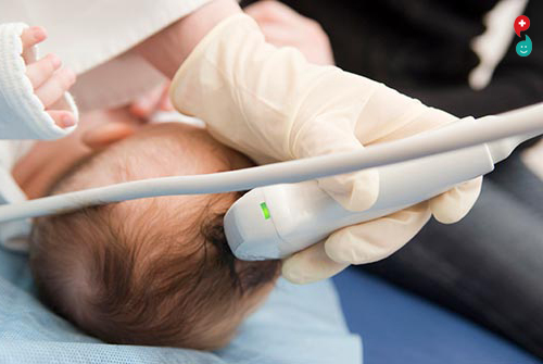
Head Ultrasound
A head ultrasound examination produces images of the brain and the cerebrospinal fluid that flows and is contained within its ventricles, the fluid filled cavities located in the deep portion of the brain. Since ultrasound waves do not pass through bone easily, this exam is most commonly performed on infants, whose skulls have not completely formed. The gaps between those skull bones provide a "window," allowing the ultrasound beam to freely pass into and back from the brain. The ultrasound probe and some gel are placed on the outside of the head in one of those regions without bone.
Transcranial Doppler
A transcranial Doppler (TCD) ultrasound evaluates both the direction and velocity of the blood flow in the major cerebral arteries of the brain. This type of ultrasound exam is also used during surgical procedures to monitor blood flow in the brain. TCD may be used alone or with other diagnostic exams such as magnetic resonance imaging (MRI), magnetic resonance angiography (MRA) and computed tomography (CT) scans.
Ultrasound is safe and painless, and produces pictures of the inside of the body using sound waves. Ultrasound imaging, also called ultrasound scanning or sonography, involves the use of a small transducer (probe) and ultrasound gel placed directly on the skin. High-frequency sound waves are transmitted from the probe through the gel into the body. The transducer collects the sounds that bounce back and a computer then uses those sound waves to create an image. Ultrasound examinations do not use ionizing radiation (as used in x-rays), thus there is no radiation exposure to the patient. Because ultrasound images are captured in real-time, they can show the structure and movement of the body's internal organs, as well as blood flowing through blood vessels.
Ultrasound imaging is a noninvasive medical test that helps physicians diagnose and treat medical conditions.