Overview
A fetal ultrasound (sonogram) is an imaging technique that uses sound waves to produce images of a fetus in the uterus. Fetal ultrasound images can help your health care provider evaluate your baby's growth and development and monitor your pregnancy. In some cases, fetal ultrasound is used to evaluate possible problems or help confirm a diagnosis.
The first fetal ultrasound is usually done during the first trimester to confirm the pregnancy and estimate how long you've been pregnant. If your pregnancy remains uncomplicated, the next ultrasound is typically offered during the second trimester, when anatomic details are visible. If a problem is suspected, a follow-up ultrasound or additional imaging tests, such as an MRI, might be recommended.
There are two main types of fetal ultrasound exams:
Transvaginal ultrasound. With this type of fetal ultrasound, a wandlike device called a transducer is placed in your vagina to send out sound waves and gather the reflections. Transvaginal ultrasounds are used most often during early pregnancy. This type of ultrasound also might be done if a transabdominal ultrasound didn't provide enough information.
Transabdominal ultrasound. A transabdominal fetal ultrasound is done by moving a transducer over your abdomen.
Various other types of transabdominal ultrasounds are available, including:
Specialized sonographic evaluation. This type of exam might be needed in specific circumstances, such as when a fetal abnormality is known or suspected. In this situation, a more detailed evaluation can provide additional information about the abnormality.
3D ultrasound. This exam provides a two-dimensional display of three-dimensional data. This type of ultrasound is sometimes used to help health care providers detect facial abnormalities or neural tube defects.
Doppler ultrasound. A Doppler ultrasound measures slight changes in the ultrasound waves as they bounce off moving objects, such as blood cells. It can provide details about a baby's blood flow.
Fetal echocardiography. This exam provides a detailed picture of a baby's heart. It might be used to confirm or rule out a congenital heart defect.
Why it's done
First trimester ultrasound examination is done to evaluate the presence, size and location of the pregnancy, determine the number of fetuses, and estimate how long you've been pregnant (gestational age). Ultrasound can also be used for first trimester genetic screening, as well as screening for abnormalities of your uterus or cervix.
In the second or third trimester a standard ultrasound is done to evaluate several features of the pregnancy, including fetal anatomy. This exam is typically done between weeks 18 and 20 of pregnancy. However, the timing of this ultrasound might be altered for reasons such as obesity, which could limit visualization of the fetus.
During the second and third trimesters, limited ultrasound evaluation might be needed when a specific question requires investigation. Examples include the evaluation of fetal growth and the estimation of amniotic fluid volume. A specialized or detailed exam is done when an anomaly is suspected based on your history or other prenatal exam results.
Your health care provider might use fetal ultrasound to:
Confirm the pregnancy and its location. Some fetuses develop outside of the uterus, in the fallopian tube. A fetal ultrasound can help your health care provider detect a pregnancy outside of the uterus (ectopic pregnancy).
Determine your baby's gestational age. Knowing the baby's age can help your health care provider determine your due date and track various milestones throughout your pregnancy.
Confirm the number of babies. If your health care provider suspects a multiple pregnancy, an ultrasound might be done to confirm the number of babies.
Evaluate your baby's growth. Your health care provider can use ultrasound to determine whether your baby is growing at a normal rate. Ultrasound can be used to monitor your baby's movement, breathing and heart rate.
Study the placenta and amniotic fluid levels. The placenta provides your baby with vital nutrients and oxygen-rich blood. Too much or too little amniotic fluid the fluid that surrounds the baby in the uterus during pregnancy or complications with the placenta need special attention. An ultrasound can help evaluate the placenta and amniotic fluid around the baby.
Identify birth defects. An ultrasound can help your health care provider screen for some birth defects.
Investigate complications. If you're bleeding or having other complications, an ultrasound might help your health care provider determine the cause.
Perform other prenatal tests. Your health care provider might use ultrasound to guide needle placement during certain prenatal tests, such as amniocentesis or chorionic villus sampling.
Determine fetal position before delivery. Most babies are positioned headfirst by the end of the third trimester. That doesn't always happen, though. Ultrasound imaging can confirm the baby's presentation so that your health care provider can discuss options for delivery.
Fetal ultrasound should be done only for valid medical reasons. Fetal ultrasound isn't recommended only to determine a baby's sex. Similarly, fetal ultrasound isn't recommended solely for the purpose of producing keepsake videos or pictures.
If your health care provider doesn't suggest a fetal ultrasound but you'd like the reassurance an ultrasound can provide, share your wishes with your care provider so that you can work together to determine what's best for you and your baby.
Risks
Diagnostic ultrasound has been used during pregnancy for many years and is generally considered safe when used appropriately. The lowest amount of ultrasound energy that provides an accurate assessment should be used.
Fetal ultrasound also has limitations. Fetal ultrasound might not detect all birth defects or might incorrectly suggest a birth defect is present when it's not.
How you prepare
You might be asked to drink a certain amount of fluid or avoid urinating before a fetal ultrasound, depending on the type of ultrasound. When scheduling your ultrasound, ask your health care provider for instructions.
Also be aware that fetal ultrasound can be done through the vagina (transvaginal) or over the abdomen (transabdominal), depending on why it's being done or the stage of your pregnancy. If you're having a transabdominal ultrasound, consider wearing loosefitting clothing so that you can easily expose your abdomen.
What you can expect
During the procedure
During a transabdominal fetal ultrasound, you'll recline on an exam table and expose your abdomen. Your health care provider or technician will apply a special gel to your abdomen. This will improve the conduction of sound waves and eliminate air between your skin and the transducer.
Your health care provider or technician will move or scan the transducer back and forth over your abdomen. The sound waves reflected off your bones and other tissues will be converted into images on a monitor.
Your health care provider or technician will measure your baby's anatomy. He or she might print or store certain images to document important structures. You'll likely be given copies of some of the images.
Depending on your baby's position and stage of development, you might be able to make out a face, hands and fingers, or arms and legs. Don't worry if you can't "see" your baby. Ultrasound images can be hard for an untrained observer to decipher. Ask your health care provider or technician to explain what's on the screen.
The procedure for other types of fetal ultrasound exams is similar. If you're having a transvaginal ultrasound, however, you'll be asked to change into a hospital gown or undress from the waist down. You'll recline on an exam table and place your feet in stirrups. The transducer will be covered in a plastic sheath, like a condom, and be lubricated with gel. Your health care provider or technician will place the transducer in your vagina.
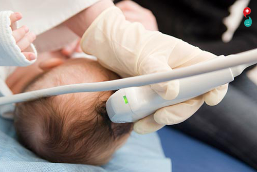
Cranial Ultrasound/Head Ultrasound
Ultrasound imaging of the head uses sound waves to produce pictures of the brain and cerebrospinal fluid. It is most commonly performed on infants, whose skulls have not completely formed. A transcranial Doppler ultrasound evaluates blood flow in the brain's major arteries. Ultrasound is safe, noninvasive, and does not use ionizing radiation.
This procedure requires little to no special preparation. Your doctor will instruct you on how to prepare, including whether adults undergoing the exam should refrain from using nicotine-based products that may cause blood vessels to constrict. Leave jewelry at home and wear loose, comfortable clothing. You may be asked to wear a gown.
What is cranial ultrasound?
Head and transcranial Doppler are two types of cranial ultrasound exams used to evaluate brain tissue and the flow of blood to the brain, respectively.
Head Ultrasound
A head ultrasound examination produces images of the brain and the cerebrospinal fluid that flows and is contained within its ventricles, the fluid filled cavities located in the deep portion of the brain. Since ultrasound waves do not pass through bone easily, this exam is most commonly performed on infants, whose skulls have not completely formed. The gaps between those skull bones provide a "window," allowing the ultrasound beam to freely pass into and back from the brain. The ultrasound probe and some gel are placed on the outside of the head in one of those regions without bone.
Transcranial Doppler
A transcranial Doppler (TCD) ultrasound evaluates both the direction and velocity of the blood flow in the major cerebral arteries of the brain. This type of ultrasound exam is also used during surgical procedures to monitor blood flow in the brain. TCD may be used alone or with other diagnostic exams such as magnetic resonance imaging (MRI), magnetic resonance angiography (MRA) and computed tomography (CT) scans.
Ultrasound is safe and painless, and produces pictures of the inside of the body using sound waves. Ultrasound imaging, also called ultrasound scanning or sonography, involves the use of a small transducer (probe) and ultrasound gel placed directly on the skin. High-frequency sound waves are transmitted from the probe through the gel into the body. The transducer collects the sounds that bounce back and a computer then uses those sound waves to create an image. Ultrasound examinations do not use ionizing radiation (as used in x-rays), thus there is no radiation exposure to the patient. Because ultrasound images are captured in real-time, they can show the structure and movement of the body's internal organs, as well as blood flowing through blood vessels.
Ultrasound imaging is a noninvasive medical test that helps physicians diagnose and treat medical conditions.

गर्भाचे अल्ट्रासाऊंड (सोनोग्राम) हे इमेजिंग तंत्र आहे जी गर्भाशयात गर्भाच्या प्रतिमा तयार करण्यासाठी ध्वनी लाटा वापरते. गर्भ अल्ट्रासाऊंड प्रतिमा आपल्या आरोग्य सेवा प्रदात्यास आपल्या मुलाच्या वाढीचे आणि विकासाचे मूल्यांकन आणि आपल्या गर्भधारणाचे परीक्षण करण्यात मदत करू शकतात. काही प्रकरणांमध्ये, गर्भ अल्ट्रासाऊंड संभाव्य समस्यांचे मूल्यांकन करण्यासाठी किंवा निदान पुष्टी करण्यात मदत करण्यासाठी वापरली जाते.
प्रथम गर्भ अल्ट्रासाऊंड सामान्यतः गर्भधारणाची पुष्टी करण्यासाठी आणि आपण गर्भवती असल्यापासून किती काळ झाला याची कल्पना करण्यासाठी पहिल्या तिमाहीत करण्यात येते. जर आपली गर्भधारणा अजिबात राहिली नाही, तर पुढील अल्ट्रासाऊंड सामान्यत: दुसऱ्या तिमाहीच्या दरम्यान दिली जाते, जेव्हा ऍनाटॉमिक तपशील दृश्यमान असतात. एखाद्या समस्येचा संशय असल्यास, फॉलो-अप अल्ट्रासाऊंड किंवा एमआरआयसारख्या अतिरिक्त इमेजिंग चाचण्यांची शिफारस केली जाऊ शकते.
भ्रूण अल्ट्रासाऊंडच्या दोन मुख्य प्रकार आहेत, ट्रान्सव्हॅग्नल अल्ट्रासाऊंड. या प्रकारच्या गर्भाच्या अल्ट्रासाऊंडसह, ध्वनी लाटा पाठविण्यासाठी आणि प्रतिबिंब गोळा करण्यासाठी आपल्या योनिमध्ये ट्रान्स्ड्यूसर नावाचे एक उपकरण ठेवले जाते. ट्रान्सव्हॅगिनल अल्ट्रासाऊंडचा वापर लवकर गर्भधारणादरम्यान केला जातो. जर ट्रांसबॅडोमिनल अल्ट्रासाऊंडने पुरेशी माहिती पुरविली नसेल तर अल्ट्रासाऊंडचा देखील हा प्रकार केला जाऊ शकतो.
ट्रांसबडोमिनल अल्ट्रासाऊंड.
ट्रान्सबाडॅमिनल फेटल अल्ट्रासाऊंड आपल्या पोटावर एक ट्रान्सड्यूसर हलवून केले जाते.
इतर प्रकारचे ट्रान्सॅबडोमिनल अल्ट्रासाऊंड उपलब्ध आहेत,
विशेषीकृत सोनोग्राफीय मूल्यांकन. अशा प्रकारच्या परीक्षेत विशिष्ट परिस्थितींमध्ये आवश्यक असू शकते, जसे की जेव्हा गर्भाच्या असामान्यता ज्ञात किंवा संशयास्पद असतात. या परिस्थितीत, अधिक तपशीलवार मूल्यांकन असामान्यतेबद्दल अतिरिक्त माहिती प्रदान करू शकते.
3 डी अल्ट्रासाऊंड ही परीक्षा त्रि-आयामी डेटाचे द्वि-आयामी प्रदर्शन प्रदान करते. या प्रकारचा अल्ट्रासाऊंड कधीकधी आरोग्य सेवा प्रदात्यांना चेहर्याचा असामान्यपणा किंवा न्यूरल ट्यूब दोष ओळखण्यात मदत करण्यासाठी वापरला जातो.
डॉपलर अल्ट्रासाऊंड. डॉपलर अल्ट्रासाऊंड अल्ट्रासाऊंड लाटामध्ये किंचित बदल मोजतात कारण ते रक्त पेशीसारख्या हलणार्या वस्तू बंद करतात. हे एखाद्या बाळाच्या रक्त प्रवाह बद्दल तपशील प्रदान करू शकते.
फेटल इकोकार्डियोग्राफी. ही परीक्षा मुलाच्या हृदयाचे तपशीलवार चित्र देते. जन्मकुंडलीच्या हृदयरोगाची पुष्टी करण्यासाठी किंवा त्यांचा निषेध करण्यासाठी याचा वापर केला जाऊ शकतो.
ते का केले
प्रथम त्रैमासिका अल्ट्रासाऊंड परीक्षा गर्भधारणेची उपस्थिति, आकार आणि स्थानाचे मूल्यांकन करण्यासाठी, गर्भाची संख्या निर्धारित करण्यासाठी आणि आपण गर्भधारणा किती काळ (गर्भावस्थेच्या वयाची) आहे याचा अंदाज घेण्यासाठी केला जातो. अल्ट्रासाऊंडचा वापर प्रथम तिमाही अनुवांशिक स्क्रीनिंग तसेच आपल्या गर्भाशयाच्या किंवा गर्भाशयाच्या विकृतींसाठी स्क्रीनिंग देखील केला जाऊ शकतो.
दुसऱ्या किंवा तिसऱ्या तिमाहीत गर्भधारणा समेत गर्भधारणेच्या अनेक वैशिष्ट्यांचे मूल्यांकन करण्यासाठी मानक अल्ट्रासाऊंड केले जाते. ही परीक्षा सामान्यत: गर्भावस्थेच्या 18 आणि 20 आठवड्यांच्या दरम्यान केली जाते. तथापि, या अल्ट्रासाऊंडचा कालावधी लठ्ठपणासारख्या कारणास्तव बदलला जाऊ शकतो, ज्यामुळे गर्भाच्या दृश्यमानतेस मर्यादा येऊ शकते.
दुसऱ्या आणि तिसऱ्या तिमाहीत दरम्यान, जेव्हा एखाद्या विशिष्ट प्रश्नाची तपासणी आवश्यक असते तेव्हा मर्यादित अल्ट्रासाऊंड मूल्यांकनाची आवश्यकता असू शकते. गर्भाच्या वाढीचे मूल्यांकन आणि अम्नीओटिक फ्लुइड व्हॉल्यूमचा अंदाज समाविष्ट आहे. आपल्या इतिहासाच्या किंवा इतर प्रसुतीपूर्व परीणामांच्या परिणामांवर आधारित विसंगतीचा संशय असल्यास विशिष्ट किंवा तपशीलवार परीक्षा केली जाते.
आपले हेल्थ केअर प्रदाता गर्भ अल्ट्रासाऊंडचा वापर करू शकतात:
गर्भधारणा आणि त्याचे स्थान याची पुष्टी करा. काही गर्भ फॅलोपियन नलिकेत गर्भाशयाच्या बाहेर विकसित होतात. गर्भाच्या अल्ट्रासाऊंडमुळे आपल्या आरोग्य सेवा प्रदात्यास गर्भाशयाच्या बाहेर गर्भधारणा शोधण्यास मदत होते (एक्टोपिक गर्भावस्था).
आपल्या बाळाच्या गर्भधारणाची वय निर्धारित करा. बाळाचे वय जाणून घेणे आपल्या आरोग्य सेवा प्रदात्यास आपली देय तारीख निर्धारित करण्यास आणि आपल्या गर्भधारणादरम्यान विविध मैलाचा मागोवा घेण्यास मदत करू शकते.
बाळांची संख्या निश्चित करा. आपल्या आरोग्य सेवा प्रदात्यास एकाधिक गर्भधारणेस शंका असल्यास, बाळांची संख्या निश्चित करण्यासाठी अल्ट्रासाऊंड केले जाऊ शकते.
आपल्या मुलाच्या वाढीचे मूल्यांकन करा. आपले बाळ सामान्य दराने वाढत आहे काय हे निर्धारित करण्यासाठी आपला आरोग्य सेवा प्रदाता अल्ट्रासाऊंड वापरू शकतो. अल्ट्रासाऊंडचा वापर आपल्या मुलाच्या हालचाली, श्वासोच्छवासाचे आणि हृदयविकाराचे निरीक्षण करण्यासाठी केला जाऊ शकतो.
प्लेसेंटा आणि अम्नीओटिक द्रव पातळीचा अभ्यास करा. प्लेसेंटा आपल्या बाळाला पोषक तत्त्वे आणि ऑक्सिजन समृध्द रक्त पुरवतो. गर्भधारणेदरम्यान गर्भाशयात बाळाच्या आसपास असलेल्या द्रवपदार्थात खूप जास्त किंवा खूपच कमी अम्नीओटिक द्रव - किंवा प्लेसेंटासह गुंतागुंतांना विशेष लक्ष देणे आवश्यक आहे. अल्ट्रासाऊंड बाळाच्या आसपास प्लेसेंटा आणि अम्नीओटिक द्रव्यांचे मूल्यांकन करण्यात मदत करू शकते.
जन्म दोष ओळखणे. अल्ट्रासाऊंड आपल्या आरोग्य सेवा प्रदात्यास काही जन्माच्या दोषांसाठी मदत करू शकते.
गुंतागुंत तपासा. आपण रक्तस्त्राव करत असल्यास किंवा इतर गुंतागुंत असल्यास, अल्ट्रासाऊंड आपल्या आरोग्य सेवा प्रदात्यास कारणीभूत ठरविण्यात मदत करू शकते.
इतर जन्मपूर्व चाचण्या करा. आपल्या आरोग्य सेवा प्रदात्याने अल्ट्रासाऊंडचा वापर काही विशिष्ट प्रसवपूर्व चाचण्यांमध्ये सूक्ष्म प्लेसमेंटच्या मार्गदर्शनासाठी करू शकता जसे की अॅनिनिसेनेसिस किंवा कोरियोनिक विल्स सॅम्पलिंग.
वितरण करण्यापूर्वी गर्भाची स्थिती निर्धारित करा. तिसऱ्या तिमाहीच्या शेवटी बहुतेक बाळांना मुख्य परीक्षेत स्थान दिले जाते. ते नेहमी होत नाही तरी. अल्ट्रासाऊंड इमेजिंग मुलाच्या प्रेझेंटेशनची पुष्टी करू शकते जेणेकरुन आपले हेल्थ केअर प्रदाता वितरणासाठी पर्याय चर्चा करू शकेल.
गर्भाशयाचे अल्ट्रासाऊंड फक्त वैद्यकीय कारणांसाठीच केले पाहिजे. गर्भाशयाचे अल्ट्रासाऊंड केवळ बाळाच्या सेक्सचे निर्धारण करण्याची शिफारस केलेली नाही. त्याचप्रमाणे, लेटल अल्ट्रासाऊंडला केवळ देस्टेक व्हिडिओ किंवा चित्रे तयार करण्याच्या उद्देशाने शिफारस केली जात नाही.
जर आपले हेल्थ केअर प्रदाता गर्भाच्या अल्ट्रासाऊंडचा सल्ला देत नाही तर आपल्याला अल्ट्रासाऊंड देऊ शकेल अशी आश्वासन आपल्याला आवडेल, आपल्या इच्छा प्रदात्यासह आपली इच्छा सामायिक करा जेणेकरुन आपण आणि आपल्या मुलासाठी काय सर्वोत्कृष्ट आहे हे ठरविण्यासाठी आपण एकत्र काम करू शकता.
धोके
डायग्नोस्टिक अल्ट्रासाऊंडचा वापर गर्भधारणेदरम्यान बर्याच वर्षांपासून केला जातो आणि योग्य वेळी वापरल्यास सामान्यपणे सुरक्षित मानले जाते. अचूक प्रमाण प्रदान करणारे कमीतकमी अल्ट्रासाऊंड ऊर्जा वापरली पाहिजे.
गर्भाशयाच्या अल्ट्रासाऊंडमध्ये मर्यादा देखील आहेत. गर्भाशयाचे अल्ट्रासाऊंड सर्व जन्माच्या दोषांचा शोध घेऊ शकत नाही - किंवा नसल्यास जन्म दोष हा चुकीचा असल्याचे कदाचित सूचित करेल.
आपण कसे तयार आहात?
अल्ट्रासाऊंडच्या प्रकारावर अवलंबून गर्भाच्या अल्ट्रासाऊंडच्या आधी आपल्याला विशिष्ट प्रमाणात द्रव पिण्याची किंवा मूत्रपिंड टाळण्यास सांगितले जाऊ शकते. आपले अल्ट्रासाऊंड शेड्यूल करताना, आपल्या आरोग्य सेवा प्रदात्यास निर्देशांसाठी विचारा.
हे देखील लक्षात घ्या की गर्भ अल्ट्रासाऊंड योनी मधून (ट्रान्सव्हॅगिनल) किंवा ओटीपोटा(ट्रान्सॅबडोमिनल) द्वारे केले जाऊ शकते, हे का केले जात आहे किंवा आपल्या गर्भधारणाची स्थिती यावर अवलंबून आहे. जर आपणास ट्रान्सबाडॅमिनल अल्ट्रासाऊंड येत असेल तर सैल कपडे घालण्याचा विचार करा जेणेकरून आपण सहजपणे आपल्या पोटास उघडकीस आणू शकता.
आपण काय अपेक्षा करू शकता?
प्रक्रिया दरम्यान
ट्रान्सबाडॅमिनल फेटल अल्ट्रासाऊंड दरम्यान, आपण परीक्षा टेबलावर रेखांकित कराल आणि आपले ओटीपोट उघडेल. आपल्या आरोग्यासाठी प्रदाता किंवा तंत्रज्ञ आपल्या पोटात एक विशेष जेल लागू करतील. यामुळे ध्वनी लाटा चालवल्या जातील आणि आपल्या त्वचे आणि ट्रान्सड्यूसर दरम्यान हवा दूर होईल.
आपला आरोग्य सेवा प्रदाता किंवा तंत्रज्ञ आपल्या पेटीवर पुढे आणि पुढे ट्रान्सड्यूसर स्कॅन करेल किंवा स्कॅन करेल. आपल्या हाडे आणि इतर उतींना परावर्तित केलेल्या ध्वनी लाटा मॉनिटरवर प्रतिमांमध्ये रूपांतरित केल्या जातील.
आपला आरोग्य सेवा प्रदाता किंवा तंत्रज्ञ आपल्या बाळाच्या शरीर रचनांचा मापन करेल. ठराविक संरचना दस्तावेज करण्यासाठी तो किंवा ती काही प्रतिमा मुद्रित किंवा संग्रहित करू शकेल. आपल्याला कदाचित काही प्रतिमांची प्रतिलिपी दिली जाईल.
आपल्या मुलाच्या स्थिती आणि विकासाच्या चरणावर अवलंबून, आपण चेहरा, हात आणि बोटांनी किंवा हात व पाय काढण्यास सक्षम असाल. आपण आपल्या बाळाला "पाहू" शकत नसल्यास काळजी करू नका. अनावृत्त प्रेक्षकांना समजण्यासाठी अल्ट्रासाऊंड प्रतिमा कठिण असू शकतात. स्क्रीनवर काय आहे हे स्पष्ट करण्यासाठी आपल्या आरोग्य सेवा प्रदाता किंवा तंत्रज्ञानास विचारा.
इतर प्रकारच्या गर्भाच्या अल्ट्रासाऊंड परीक्षणाची प्रक्रिया समान आहे. जर आपल्याला ट्रान्सव्हॅग्नल अल्ट्रासाऊंड येत असेल तर, आपल्याला हॉस्पिटल गाउनमध्ये बदलण्याची किंवा कमर खाली उतरविण्यास सांगितले जाईल. आपण परीक्षेच्या टेबलावर रेखांकित कराल आणि आपले पाय ररक्यांना ठेवतील. ट्रान्सड्यूसरला प्लास्टिकच्या म्यानमध्ये, कंडोमप्रमाणे ढकलले जाईल आणि जेलने स्नेही केले जाईल. आपला आरोग्य सेवा प्रदाता किंवा तंत्रज्ञ आपल्या योनिमध्ये ट्रान्सड्यूसर ठेवेल.

क्रेनियल अल्ट्रासाऊंड / डोके अल्ट्रासाऊंड :
डोकेच्या अल्ट्रासाऊंड इमेजिंगमुळे मेंदू आणि सेरेब्रोस्पिनील द्रवपदार्थांच्या चित्र तयार करण्यासाठी ध्वनी लाटा वापरतात. हे सर्वसाधारणपणे शिशुंवर केले जाते, त्यांचे खोके पूर्णतः तयार झालेले नाहीत. ट्रान्सक्रॅनलियल डॉपलर अल्ट्रासाऊंड मेंदूच्या मुख्य धमन्यांमध्ये रक्त प्रवाहाचे मूल्यांकन करते. अल्ट्रासाऊंड सुरक्षित, अनिश्चित आहे आणि आयओनिंग विकिरण वापरत नाही.
या प्रक्रियेस विशेष तयारीची आवश्यकता नाही. रक्ताच्या वाहनांना धक्का देण्यास कारणीभूत असलेल्या निकोटीन-आधारित उत्पादनांचा वापर करुन प्रौढांनी चाचणी करावी की नाही यासह आपले डॉक्टर आपल्याला कसे तयार करावे याबद्दल मार्गदर्शन करतील. घरी दागदागिने सोडा आणि ढीग, आरामदायक कपडे घाला. आपल्याला गाउन घालण्यास सांगितले जाऊ शकते.
कानासंबंधीचा अल्ट्रासाऊंड काय आहे?
हेड आणि ट्रान्सक्रॅनलियल डोप्लर हे दोन प्रकारचे क्रॅनियल अल्ट्रासाऊंड परीक्षा आहेत ज्याचा क्रमशः मेंदूतील ऊतक आणि रक्ताचा प्रवाह मेंदूचे मूल्यांकन करण्यासाठी केला जातो.
डोके अल्ट्रासाऊंड :
डोके अल्ट्रासाऊंड परीक्षेत मेंदूच्या प्रतिमा आणि सेरेब्रोस्पिनील द्रवपदार्थ तयार होतात जो त्याच्या वेंट्रिकल्समध्ये प्रवाहित होतो आणि मेंदूच्या खोल भागामध्ये स्थित द्रवपदार्थ असलेल्या पोकळी. अल्ट्रासाऊंड लाटा सहजपणे हाडांच्या माध्यमातून जात नाहीत म्हणून, ही परीक्षा सर्वात सामान्यपणे नवजात मुलांवर केली जाते, ज्याच्या खोपण्या पूर्णपणे तयार होत नाहीत. त्या खोपड्या हाडे दरम्यान अंतर, "खिडकी" प्रदान करते ज्यामुळे अल्ट्रासाऊंड बीम मुक्तपणे मस्तिष्कमधून व मागे जाण्याची परवानगी देते. अल्ट्रासाऊंड प्रोब आणि काही जेल हड्डीशिवाय त्यापैकी एका भागात डोकेच्या बाहेरील बाजूस ठेवले जातात.
ट्रान्सक्रॅनियल डॉपलर :
ट्रान्सक्रॅनलियल डॉपलर (टीसीडी) अल्ट्रासाऊंड मेंदूच्या मुख्य सेरेब्रल धमन्यांमध्ये रक्त प्रवाहच्या दिशेने व वेगाने दोन्हीचे मूल्यांकन करते. या प्रकारचे अल्ट्रासाऊंड परीक्षा देखील मेंदूतील रक्त प्रवाहांचे परीक्षण करण्यासाठी शस्त्रक्रिया प्रक्रियेदरम्यान वापरली जाते. टीसीडीचा वापर एकट्याने किंवा चुंबकीय अनुनाद इमेजिंग (एमआरआय), चुंबकीय अनुनाद एंजियोग्राफी (एमआरए) आणि संगणित टोमोग्राफी (सीटी) स्कॅन सारख्या इतर निदानात्मक परीक्षांसह केला जाऊ शकतो.
अल्ट्रासाऊंड सुरक्षित आणि वेदनाहीन आहे आणि ध्वनीच्या लाटा वापरून शरीराच्या आतील चित्र तयार करते. अल्ट्रासाऊंड इमेजिंग, ज्याला अल्ट्रासाऊंड स्कॅनिंग किंवा सोनोग्राफी देखील म्हटले जाते, त्यात एक लहान ट्रान्सड्यूसर (प्रोब) आणि अल्ट्रासाऊंड जेलचा वापर त्वचेवर थेट ठेवला जातो. उच्च-फ्रिक्वेंसी ध्वनी लाटा तपासणीतून जेलमधून शरीरात प्रवेश करतात. ट्रान्सड्यूसर आवाज परत आणणार्या कॉम्प्युटरला एकत्र करतो आणि नंतर एक आवाज तयार करण्यासाठी त्या ध्वनी लाटा वापरतो. अल्ट्रासाऊंड परीक्षणे आयोनायझेशन रेडिएशन (एक्स-किरणांमध्ये वापरल्याप्रमाणे) वापरत नाहीत, अशा प्रकारे रोग्यास विकिरण नाही. अल्ट्रासाऊंड प्रतिमा रीअल-टाइममध्ये कॅप्चर केल्या गेल्यामुळे ते शरीराच्या अंतर्गत अवयवांची संरचना आणि हालचाल तसेच रक्तवाहिन्यांमधून वाहणारे रक्त दर्शवू शकतात.
अल्ट्रासाऊंड इमेजिंग एक नॉनविवासिव्ह मेडिकल टेस्ट आहे जे वैद्यकीय परिस्थांचे निदान आणि उपचार करण्यास मदत करते.