Magnetic Resonance Imaging (MRI) - Body
Magnetic resonance imaging (MRI) of the body uses a powerful magnetic field, radio waves and a computer to produce detailed pictures of the inside of your body. It may be used to help diagnose or monitor treatment for a variety of conditions within the chest, abdomen and pelvis. If you're pregnant, body MRI may be used to safely monitor your baby.
Tell your doctor about any health problems, recent surgeries or allergies and whether there's a possibility you are pregnant. The magnetic field is not harmful, but it may cause some medical devices to malfunction. Most orthopedic implants pose no risk, but you should always tell the technologist if you have any devices or metal in your body. Guidelines about eating and drinking before your exam vary between facilities. Unless you are told otherwise, take your regular medications as usual. Leave jewelry at home and wear loose, comfortable clothing. You may be asked to wear a gown. If you have claustrophobia or anxiety, you may want to ask your doctor for a mild sedative prior to the exam.
What is MRI of the Body? What are some common uses of the procedure? How should I prepare for the procedure? What does the equipment look like? How does the procedure work? How is the procedure performed?
What will I experience during and after procedure? Who interprets the results and how do I get them? What are the benefits vs. risks? What are the limitations of MRI of the Body? Which test, procedure or treatment is best for me?
What is MRI of the Body?
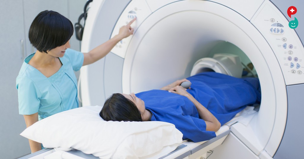
Magnetic resonance imaging (MRI) is a noninvasive medical test that physicians use to diagnose medical conditions.
MRI uses a powerful magnetic field, radio frequency pulses and a computer to produce detailed pictures of organs, soft tissues, bone and virtually all other internal body structures. MRI does not use ionizing radiation (x-rays).
Detailed MR images allow physicians to evaluate various parts of the body and determine the presence of certain diseases. The images can then be examined on a computer monitor, transmitted electronically, printed or copied to a CD or uploaded to a digital cloud server.
top of page
What are some common uses of the procedure?
MR imaging of the body is performed to evaluate:
organs of the chest and abdomenincluding the heart, liver, biliary tract, kidneys, spleen, bowel, pancreas, and adrenal glands.
pelvic organs including the bladder and the reproductive organs such as the uterus and ovaries in females and the prostate gland in males.
blood vessels (including MR Angiography).
lymph nodes.
Physicians use an MR examination to help diagnose or monitor treatment for conditions such as:
tumors of the chest, abdomen or pelvis.
diseases of the liver, such as cirrhosis, and abnormalities of the bile ducts and pancreas.
inflammatory bowel disease such as Crohn's disease and ulcerative colitis.
heart problems, such as congenital heart disease.
malformations of the blood vessels and inflammation of the vessels (vasculitis).
a fetus in the womb of a pregnant woman.
top of page
How should I prepare for the procedure?
You may be asked to wear a gown during the exam or you may be allowed to wear your own clothing if it is loose-fitting and has no metal fasteners.
Guidelines about eating and drinking before an MRI exam vary with the specific exam and with the imaging facility. Unless you are told otherwise, you may follow your regular daily routine and take food and medications as usual.
Some MRI examinations may require you to receive an injection of contrast material into the bloodstream. The radiologist, technologist or a nurse may ask if you have allergies of any kind, such as an allergy to iodine or x-ray contrast material, drugs, food, or the environment, or if you have asthma. The contrast material most commonly used for an MRI exam contains a metal called gadolinium. Gadolinium can be used in patients with iodine contrast allergy. It is far less common for a patient to have an allergy to a gadolinium-based contrast agent used for MRI than the iodine-containing contrast for CT. However, even if it is known that the patient has an allergy to the gadolinium contrast, it may still be possible to use it after appropriate pre-medication. Patient consent will be requested in this instance. For more information on adverse reactions to gadolinium-based contrast agents, please consult the ACR Manual on Contrast Media.
You should also let the radiologist know if you have any serious health problems, or if you have had any recent surgeries. Some conditions, such as severe kidney disease, may prevent you from being given gadolinium contrast for an MRI. If you have a history of kidney disease or liver transplant, it will be necessary to perform a blood test to determine whether the kidneys are functioning adequately.
Women should always inform their physician or technologist if there is any possibility that they are pregnant. MRI has been used for scanning patients since the 1980s with no reports of any ill effects on pregnant women or their unborn babies. However, because the unborn baby will be in a strong magnetic field, pregnant women should not have this exam in the first three to four months of pregnancy unless the potential benefit from the MRI exam is assumed to outweigh the potential risks. Pregnant women should not receive injections of gadolinium contrast material except when absolutely necessary for medical treatment. See the MRI Safety page for more information about pregnancy and MRI.
If you have claustrophobia (fear of enclosed spaces) or anxiety, you may want to ask your physician for a prescription for a mild sedative prior to your scheduled examination.
Jewelry and other accessories should be left at home, if possible, or removed prior to the MRI scan. Because they can interfere with the magnetic field of the MRI unit, metal and electronic items are not allowed in the exam room. In addition to affecting the MRI images, these objects can become projectiles within the MRI scanner room and may cause you and/or others nearby harm. These items include:
jewelry, watches, credit cards and hearing aids, all of which can be damaged
pins, hairpins, metal zippers and similar metallic items, which can distort MRI images
removable dental work
pens, pocket knives and eyeglasses
body piercings
In most cases, an MRI exam is safe for patients with metal implants, except for a few types. People with the following implants cannot be scanned and should not enter the MRI scanning area:
cochlear (ear) implant
some types of clips used for brain aneurysms
some types of metal coils placed within blood vessels
nearly all cardiac defibrillators and pacemakers
You should tell the technologist if you have medical or electronic devices in your body. These objects may interfere with the exam or potentially pose a risk, depending on their nature and the strength of the MRI magnet. Many implanted devices will have a pamphlet explaining the MRI risks for that particular device. If you have the pamphlet, it is useful to bring that to the attention of the scheduler before the exam and bring it to your exam in case the radiologist or technologist has any questions. Some implanted devices require a short period of time after placement (usually six weeks) before being safe for MRI examinations. Examples include but are not limited to:
artificial heart valves
implanted drug infusion ports
artificial limbs or metallic joint prostheses
implanted nerve stimulators
metal pins, screws, plates, stents or surgical staples
If there is any question of their presence, an x-ray may be taken to detect and identify any metal objects. In general, metal objects used in orthopedic surgery pose no risk during MRI. However, a recently placed artificial joint may require the use of another imaging procedure.
Patients who might have metal objects in certain parts of their bodies may also require an x-ray prior to an MRI. You should notify the technologist or radiologist of any shrapnel, bullets, or other pieces of metal that may be present in your body due to prior accidents. Foreign bodies near and especially lodged in the eyes are particularly important because they may move during the scan, possibly causing blindness. Dyes used in tattoos may contain iron and could heat up during an MRI scan, but this is rare. Tooth fillings and braces usually are not affected by the magnetic field, but they may distort images of the facial area or brain, so you should let the radiologist know about them.

Knee MRI Test
A knee MRI (magnetic resonance imaging) scan uses energy from strong magnets to create pictures of the knee joint and muscles and tissues.An MRI does not use radiation (x-rays). Single MRI images are called slices. The images can be stored on a computer or printed on film. One exam produces many images.
How the Test is Performed
You will wear a hospital gown or clothes without metal zippers or snaps (such as sweatpants and a t-shirt). Please remove your watches, jewelry, and wallet. Certain types of metal can cause blurry images.You will lie on a narrow table that slides into a large tunnel-like scanner.
Some exams use a special dye (contrast). Most of the time, you will get the dye through a vein (IV) in your arm or hand before the test. Sometimes, the dye is injected into a joint. The dye helps the radiologist see certain areas more clearly.During the MRI, the person who operates the machine will watch you from another room. The test most often lasts 30 to 60 minutes, but may take longer. It can be loud. The technician can give you some ear plugs if needed.
How to Prepare for the Test
You may be asked not to eat or drink anything for 4 to 6 hours before the scan.
Tell your health care provider if you are afraid of closed spaces (have claustrophobia). You may be given a medicine to help you feel sleepy and less anxious. Your provider may suggest an "open" MRI, in which the machine is not as close to the body.
Before the test, tell your provider if you have:
-Brain aneurysm clips
-Certain types of artificial heart valves
-Heart defibrillator or pacemaker
-Inner ear (cochlear) implants
-Kidney disease or dialysis (you may not be able to receive contrast)
-Recently placed artificial joints
-Certain types of vascular stents
-Worked with sheet metal in the past (you may need tests to check for metal pieces in your eyes)
Because the MRI contains strong magnets, metal objects are not allowed into the room with the MRI scanner:
-Pens, pocketknives, and eyeglasses may fly across the room.
-Items such as jewelry, watches, credit cards, and hearing aids can be damaged.
-Pins, hairpins, metal zippers, and similar metallic items can distort the images.
-Removable dental work should be taken out just before the scan.
How the Test will Feel
An MRI exam causes no pain. You will need to lie still. Too much movement can blur MRI images and cause errors.The table may be hard or cold, but you can ask for a blanket or pillow. The machine makes loud thumping and humming noises when turned on. You can wear ear plugs to help block out the noise.
An intercom in the room allows you to speak to someone at any time. Some MRIs have televisions and special headphones to help the time pass.There is no recovery time, unless you were given a medicine to relax. After an MRI scan, you can return to your normal diet, activity, and medicines.
Why the Test is Performed
Your provider may order this test if you have:
-An abnormal result on a knee x-ray or bone scan
-A feeling that your knee is giving away in the knee joint
-Buildup of joint fluid behind the knee (Baker cyst)
-Fluid collecting in the knee joint
-Infection of the knee joint
-Knee cap injury
-Knee pain with fever
-Knee locking when you walk or moving
-Signs of damage to the knee muscle, cartilage, or ligaments
-Knee pain that does not get better with treatment
-Instability of the knee
You may also have this test to check your progress after knee surgery.
Normal Results
A normal result means your knee looks OK.
What Abnormal Results Mean
Abnormal results may be due to a sprain or tear of the ligaments in the knee area.
Abnormal results may also be due to:
-Meniscus or cartilage injuries
-Arthritis of the knee
-Avascular necrosis (also called osteonecrosis)
-Bone tumor or cancer
-Broken bone
-Buildup of joint fluid behind the knee (Baker cyst)
-Infection in the bone (osteomyelitis)
-Inflammation
-Injury of the knee cap
Talk to your provider if you have questions or concerns.
Risks
MRI contains no radiation. There have been no reported side effects from the magnetic fields and radio waves.
The most common type of contrast (dye) used is gadolinium. It is very safe. Allergic reactions to the substance are rare. However, gadolinium can be harmful to people with kidney problems that need dialysis. If you have kidney problems, please tell your provider before the test.
The strong magnetic fields created during an MRI can cause heart pacemakers and other implants to not work as well. It can also cause small pieces of metal inside your body to move or shift. For safety reasons, please do not bring anything that contains metal into the scanner room.

Sitting for too long may increase the risk of dementia in middle-aged adults, according to scientists, including one of Indian origin.
In a study published in the journal PLOS One, the researchers recruited 35 people (aged between 45 and 75 years).
The researchers, including Prabha Siddarth from the University of California, Los Angeles in the US, asked the participants about their physical activity levels and the average number of hours per day they spent sitting over the previous week.
Each person had a high-resolution MRI scan, which provides a detailed look at the medial temporal lobe, or MTL, a brain region involved in the formation of new memories.
The researchers found that sedentary behaviour is a significant predictor of thinning of the MTL and that physical activity, even at high levels, is insufficient to offset the harmful effects of sitting for extended periods.
MTL thinning can be a precursor to cognitive decline and dementia in middle-aged and older adults.
Reducing sedentary behaviour may be a possible target for interventions designed to improve brain health in people at risk for Alzheimer's disease, said Siddarth.
This study does not prove that too much sitting causes thinner brain structures, but instead that more hours spent sitting are associated with thinner regions, he said.
मॅग्नेटिक रेझोनान्स इमेजिंग (एमआरआय) - बॉडी
शरीराच्या आतील तपशीलवार चित्रे तयार करण्यासाठी शरीराच्या चुंबकीय अनुनाद इमेजिंग (एमआरआय) शक्तिशाली चुंबकीय क्षेत्र, रेडिओ लहरी आणि संगणक वापरतात. छाती, उदर आणि श्रोणीच्या विविध परिस्थितींसाठी उपचारांचे निदान किंवा नियंत्रण करण्यात मदत करण्यासाठी याचा वापर केला जाऊ शकतो. आपण गर्भवती असल्यास, आपल्या बाळाचे सुरक्षितपणे निरीक्षण करण्यासाठी शरीराच्या एमआरआयचा वापर केला जाऊ शकतो.
आपल्या डॉक्टरांना कोणत्याही आरोग्यविषयक समस्येबद्दल, अलीकडील शस्त्रक्रिया किंवा एलर्जी आणि आपण गर्भवती असल्याची शक्यता असल्याबाबत सांगा. चुंबकीय क्षेत्र हानिकारक नाही, परंतु यामुळे काही वैद्यकीय उपकरण खराब होण्यास कारणीभूत ठरू शकतात. बहुतेक ऑर्थोपेडिक इम्प्लांटसना धोका नाही, परंतु आपल्याकडे आपल्या शरीरात कोणतेही डिव्हाइस किंवा धातू असल्यास आपण नेहमीच तंत्रज्ञानास सांगावे. आपल्या परीक्षांपूर्वी खाण्याच्या आणि पिण्याच्या बद्दल मार्गदर्शकतत्त्वे सुविधांदरम्यान बदलू शकतात. आपल्याला अन्यथा सांगितले जात नाही तोपर्यंत, नेहमीप्रमाणे आपली नियमित औषधे घ्या. घरी दागदागिने सोडा आणि ढीग, आरामदायक कपडे घाला. आपल्याला गाउन घालण्यास सांगितले जाऊ शकते. आपल्याला क्लॉस्ट्रोफोबिया किंवा चिंता असल्यास, आपण आपल्या डॉक्टरांना चाचणीपूर्वी सौम्य शीतलवादासाठी विचारू शकता.
शरीराच्या एमआरआय म्हणजे काय? प्रक्रियेच्या काही सामान्य वापर काय आहेत? मी प्रक्रिया कशी तयार करावी? उपकरणे कशासारखे दिसतात? प्रक्रिया कशी कार्य करते? प्रक्रिया कशी केली जाते?
प्रक्रिया दरम्यान आणि नंतर मला काय अनुभव येईल? कोण परिणामांची व्याख्या करते आणि मी ते कसे प्राप्त करू? बनाम जोखीम काय आहेत? शरीराच्या एमआरआयची मर्यादा काय आहेत? कोणती चाचणी, प्रक्रिया किंवा उपचार माझ्यासाठी सर्वोत्तम आहे?
शरीराच्या एमआरआय म्हणजे काय?
मॅग्नेटिक रेझोनान्स इमेजिंग (एमआरआय) एक गैर-वैद्यकीय वैद्यकीय तपासणी आहे जे वैद्यकीय वैद्यकीय परिस्थितींचे निदान करण्यासाठी वापरतात.
एमआरआय एक शक्तिशाली चुंबकीय क्षेत्र, रेडिओ फ्रीक्वेंसी डाल्स आणि कॉम्प्यूटरचा वापर अंग, सॉफ्ट टिश्यूज, हाडे आणि इतर सर्व आंतरिक शरीराचे विस्तृत तपशील तयार करण्यासाठी करतात. एमआरआय आयोनायझेशन किरण (एक्स-किरण) वापरत नाही.
विस्तृत एमआर प्रतिमा चिकित्सकांना शरीराच्या विविध भागांचे मूल्यांकन करण्यासाठी आणि विशिष्ट रोगांची उपस्थिती निर्धारित करण्याची परवानगी देतात. या नंतर प्रतिमा कॉम्प्यूटर मॉनीटरवर तपासल्या जाऊ शकतात, इलेक्ट्रॉनिक पद्धतीने प्रसारित केली जाऊ शकते, सीडीवर मुद्रित केली किंवा कॉपी केली जाऊ शकते किंवा डिजिटल क्लाउड सर्व्हरवर अपलोड केली जाऊ शकते.
पृष्ठाच्या शीर्षस्थानी
प्रक्रियेच्या काही सामान्य वापर काय आहेत?
शरीराचे एमआर इमेजिंग मूल्यांकन करण्यासाठी केले जाते:
छाती आणि पोटातील अवयव-हृदय, यकृत, पित्तविषयक मार्ग, मूत्रपिंड, प्लीहा, आंत्र, आतडे आणि एड्रेनल ग्रंथी यांचा समावेश होतो.
मूत्राशयासह मूत्रपिंड आणि स्त्रियांमध्ये गर्भाशयात व अंडाशया आणि पुनरुत्पादक अवयवांचा समावेश होतो.
रक्तवाहिन्या (एमआर एंजियोग्राफीसह).
लसिका गाठी.
डॉक्टरांनी एम.आय. परीक्षा वापरली आहे ज्यायोगे यासारख्या शर्तींच्या निदानाचे निदान करण्यात किंवा त्यावर नियंत्रण ठेवण्यात मदत होईल:
छाती, उदर किंवा श्रोणि च्या अर्बुदे.
सिरोसिस सारख्या यकृताचे रोग आणि पित्त नलिका आणि पॅनक्रियाच्या असामान्यता.
क्रॉन रोग आणि अल्सरेटिव्ह कोलायटिस यासारख्या दाहक आंत्र रोग.
जन्मजात हृदय रोग जसे हृदयरोग.
रक्तवाहिन्यांच्या विकृती आणि वाहनांचा जळजळ (व्हॅस्क्युलाइटिस).
गर्भवती महिलेच्या गर्भाशयात एक गर्भ.
पृष्ठाच्या शीर्षस्थानी
मी प्रक्रिया कशी तयार करावी?
परीक्षेदरम्यान आपल्याला गाउन घालण्यास सांगितले जाऊ शकते किंवा आपल्या स्वत: च्या कपड्यांना कपडे घालण्याची परवानगी दिली जाऊ शकते आणि तिच्यात मेटल फास्टनर्स नसतील.
एमआरआय परीक्षेत आधी खाणे आणि पिणे बद्दल मार्गदर्शन विशिष्ट परीक्षेत आणि इमेजिंग सुविधासह बदलते. आपल्याला अन्यथा सांगितले जात नाही तोपर्यंत आपण आपल्या नियमित दैनंदिन नियमाचे अनुसरण करू शकता आणि नेहमीप्रमाणे अन्न व औषधे घेऊ शकता.
काही एमआरआय परीक्षा आपल्याला रक्तप्रवाहात तीव्रता सामग्रीच्या इंजेक्शन प्राप्त करण्याची आवश्यकता असू शकतात. रेडियोलॉजिस्ट, तंत्रज्ञ किंवा नर्स आयोडीन किंवा एक्स-रे कॉन्ट्रास्ट सामग्री, औषधे, अन्न किंवा वातावरणातील ऍलर्जी किंवा आपल्याला दमा असल्यास कोणत्याही प्रकारची अॅलर्जी असल्यास विचारू शकते. एमआरआय परीक्षेसाठी वापरल्या जाणार्या कॉन्ट्रास्ट सामग्रीमध्ये गॅडोलिनियम नामक धातू असते. आयोडीन कॉन्ट्रास्ट ऍलर्जी असलेल्या रुग्णांमध्ये Gadolinium वापरले जाऊ शकते. रुग्णांकडे एमडीआयसाठी वापरल्या जाणार्या गॅडोलिनियम-आधारित कॉन्ट्रास्ट एजंटची एलर्जी असणे हे फारच सामान्य आहे कारण सीओसाठी आयोडीन युक्त असलेले कॉन्ट्रास्ट. तथापि, जरी ज्ञात असले की रुग्णाला गॅडोलिनियम कॉन्ट्रास्टची ऍलर्जी आहे तरीही, योग्य पूर्व-औषधोपचारानंतर हे वापरणे अद्याप शक्य आहे. या प्रकरणात रुग्णांच्या संमतीची विनंती केली जाईल. गॅडोलिनियम-आधारित कॉन्ट्रास्ट एजंटला प्रतिकूल प्रतिक्रियांच्या अधिक माहितीसाठी, कृपया कॉन्ट्रास्ट मीडियावरील एसीआर मॅन्युअलचा सल्ला घ्या.
आपल्याला कोणतीही गंभीर आरोग्य समस्या असल्यास किंवा आपल्याकडे कोणतीही अलीकडील शस्त्रक्रिया असल्यास ती रेडियोलॉजिस्टला देखील कळवावी. गंभीर किडनी रोगासारख्या काही अटी आपल्याला एमआरआयसाठी गॅडोलिनियम कॉन्ट्रास्ट दिल्यापासून प्रतिबंधित करतात. मूत्रपिंड रोग किंवा लिव्हर ट्रान्सप्लांटचा इतिहास असल्यास, मूत्रपिंड पुरेसे कार्य करत आहेत किंवा नाही हे निर्धारित करण्यासाठी रक्त चाचणी करणे आवश्यक आहे.

गुडघा एमआरआय चाचणी :
गुडघा एमआयआय (चुंबकीय अनुनाद इमेजिंग) स्कॅन मजबूत गुंबदांमधून ऊर्जा वापरते आणि गुडघा संयुक्त आणि स्नायूंचे चित्र तयार करण्यासाठी. एक एमआरआय किरणे (एक्स-किरण) वापरत नाही. एकल एमआरआय प्रतिमा स्लाइस म्हणतात. प्रतिमा संगणकावर किंवा फिल्मवर मुद्रित केल्या जाऊ शकतात. एक परीक्षा अनेक प्रतिमा तयार करते.
चाचणी कशी केली जाते?
आपण मेटल झिप्पर किंवा स्नॅप्स (जसे sweatpants आणि t-shirt) शिवाय हॉस्पिटल गाउन किंवा कपडे घालाल. कृपया आपले घड्याळे, दागदागिने आणि वॉलेट काढून टाका. काही प्रकारचे धातू अस्पष्ट प्रतिमांचे कारण बनू शकतात. आपण एका अरुंद सारणीवर उभे राहाल जे मोठ्या सुर्यासारख्या स्कॅनरमध्ये स्लाइड करेल. काही परीक्षा विशेष रंगाचा (कॉन्ट्रास्ट) वापरतात. बर्याच वेळा, आपण चाचणीपूर्वी आपल्या हाताने किंवा हातातील शिरा (चतुर्थांश) द्वारे डाई प्राप्त कराल. कधीकधी, डाई संयुक्त मध्ये इंजेक्शन केला जातो. डाई रेडिओलॉजिस्टला काही विशिष्ट क्षेत्रे अधिक स्पष्टपणे पाहण्यास मदत करते. एमआरआयच्या दरम्यान, मशीन चालविणारी व्यक्ती आपल्याला दुसर्या खोलीत पाहतील. चाचणी बहुतेकदा 30 ते 60 मिनिटे टिकते, परंतु जास्त वेळ लागू शकतो. ते मोठ्याने असू शकते. आवश्यक असल्यास तांत्रिक आपल्याला काही कान प्लग देऊ शकेल.
चाचणीसाठी कसे तयार करावे?
- आपल्याला स्कॅनपूर्वी 4 ते 6 तास काहीही खाण्याची किंवा पिण्यास नकारण्यास सांगितले जाऊ शकते.
- आपल्याला बंद जागा (क्लॉस्ट्रोफोबिया असल्यास) घाबरल्यास आपल्याला आपले आरोग्य सेवा प्रदाता सांगा. आपल्याला झोपेचा आणि कमी उत्सुकता अनुभवण्यास मदत करण्यासाठी - आपल्याला औषध दिले जाऊ शकते. आपला प्रदाता "ओपन" एमआरआय सुचवू शकतो, ज्यामध्ये मशीन शरीराच्या जवळ नसते.
चाचणीपूर्वी, आपल्या प्रदाताला सांगा की आपल्याकडे हे आहे:
- ब्रेन एन्युरीझम क्लिप
- प्रकारचे कृत्रिम हृदय वाल्व
- हार्ट डिफिब्रिलेटर किंवा पेसमेकर
- अन्य कान (कोचलेर) रोपण
- किडनी रोग किंवा डायलिसिस (आपण कॉन्ट्रास्ट प्राप्त करण्यास सक्षम नसाल)
- रुपाने कृत्रिम जोड्या ठेवली
- वाहिन्या प्रकारचे विशिष्ट प्रकार
- भूतकाळातील शीट मेटलसह कार्य केले (आपल्या डोळ्यातील धातूचे तुकडे तपासण्यासाठी आपल्याला टेस्टची आवश्यकता असू शकते)
एमआरआयमध्ये मजबूत चुंबक असतात, एमआरआय स्कॅनरसह खोलीमध्ये धातुच्या वस्तूंना परवानगी नाही:
- पेंस, पॉकेटकेव्ह आणि चष्मा खोलीत उडतात.
- दागदागिने, घड्याळे, क्रेडिट कार्ड आणि ऐकण्याच्या सहाय्यासारख्या गोष्टी नुकसान होऊ शकतात.
- पिन, केसपिन, धातू झिपर्स, आणि अशाच धातूच्या वस्तू प्रतिमा विकृत करू शकतात.
- सक्षम करण्यायोग्य दंत काम केवळ स्कॅनपूर्वीच काढून घ्यावे.
चाचणी कशी अनुभवेल?
एमआरआय परीक्षा कोणताही त्रास होत नाही. तुला अजूनही खोटे बोलण्याची गरज आहे. खूप जास्त हालचाल एमआरआय प्रतिमा अस्पष्ट करू शकते आणि त्रुटी निर्माण करू शकते. सारणी कठोर किंवा थंड असू शकते, परंतु आपण एक कंबल किंवा उशी मागू शकता. चालू असताना मशीन जोरदार थांबायला आणि हिंगिंग आवाज बनवते. आवाज बंद करण्यास मदत करण्यासाठी आपण कान प्लग वापरू शकता.
खोलीत एक इंटरकॉम आपल्याला कोणत्याही वेळी कोणाशी तरी बोलण्याची परवानगी देतो. काही एमआरआयमध्ये वेळ निघून जाण्यासाठी टेलिव्हिजन आणि विशेष हेडफोन आहेत. आपल्याला आराम करण्यासाठी औषधे दिली जात नाही तोपर्यंत पुनर्प्राप्तीचा वेळ नाही. एमआरआय स्कॅननंतर, आपण आपल्या सामान्य आहारातील, क्रियाकलापांवर आणि औषधे परत येऊ शकता.
चाचणी का केली जाते?
आपल्या प्रदात्याने या चाचणीचे ऑर्डर आपल्यास केल्यासः
- गुडघा एक्स-रे किंवा हाड स्कॅनवर असामान्य परिणाम.
- आपल्या गुडघे गुडघा संयुक्त मध्ये देत आहे असे वाटत आहे.
- गुडघा (बॅकर सिस्ट) मागे संयुक्त द्रवपदार्थ तयार करणे.
- गुडघा संयुक्त एकत्रीत फ्लुइड.
- गुडघा संयुक्त च्या संक्रमण.
- केनी टोपी दुखापत.
- तापाने वेदना.
- आपण चालताना किंवा हलताना किक लॉकिंग करा.
- गुडघा स्नायू, उपास्थि किंवा अस्थिबंधकांना होणारे नुकसान.
- घोटीचा वेदना जो उपचाराने चांगला होत नाही.
- गुडघा च्या स्थायित्व.
- गुडघा शस्त्रक्रियेनंतर आपली प्रगती तपासण्यासाठी आपल्याकडे ही चाचणी देखील असू शकते.
सामान्य परिणाम:
सामान्य परिणाम म्हणजे आपले गुडघा ठीक दिसत आहे.
असामान्य परिणाम म्हणजे काय?
घुटमळलेल्या भागात स्नायूंच्या अस्थि किंवा अश्रूमुळे असामान्य परिणाम होऊ शकतात.
असामान्य परिणामदेखील असू शकतात:
- मनिस्कस किंवा उपास्थि जखम
- गुडघा च्या संधिशोथा
- अवास्कुलर नेक्रोसिस (ऑस्टोनोक्रोसिस देखील म्हणतात)
- बोन ट्यूमर किंवा कर्करोग
- तुटलेले हाड
- गुडघा (बॅकर सिस्ट) मागे संयुक्त द्रवपदार्थ तयार करणे
- हाड (ओस्टियोमियालाइटिस) मध्ये संक्रमण
- गुडघा कॅप च्या इजाजत
आपल्याकडे काही प्रश्न असल्यास किंवा समस्या असल्यास आपल्या प्रदात्याशी बोला.
धोके :
एमआरआयमध्ये कोणतेही विकिरण नसते. चुंबकीय क्षेत्रे आणि रेडिओ वेव्ह्सवरून कोणतेही दुष्परिणाम आढळले नाहीत.
वापरलेले सर्वात सामान्य प्रकारचे कॉन्ट्रास्ट (डाई) गॅडोलिनियम आहे. हे खूप सुरक्षित आहे. पदार्थांवरील ऍलर्जीक प्रतिक्रिया दुर्मिळ आहेत. तथापि, डायडिसिस आवश्यक असलेल्या मूत्रपिंडाच्या समस्या असलेल्या लोकांसाठी गॅडोलिनियम हानिकारक असू शकते. आपल्याला मूत्रपिंड समस्या असल्यास, कृपया चाचणीपूर्वी आपल्या प्रदात्यास सांगा.
एमआरआय दरम्यान तयार झालेले मजबूत चुंबकीय क्षेत्र हृदयाचा पेमेमेकर्स आणि इतर रोपण देखील कार्य करू शकत नाहीत. आपल्या शरीराच्या आतल्या धातूचे लहान तुकडे हलवू किंवा शिफ्ट करू शकतात. सुरक्षा कारणांमुळे, स्कॅनर रुममध्ये धातू असलेली कोणतीही वस्तू आणू नका.